Magnesium in PDB 3ohm: Crystal Structure of Activated G Alpha Q Bound to Its Effector Phospholipase C Beta 3
Enzymatic activity of Crystal Structure of Activated G Alpha Q Bound to Its Effector Phospholipase C Beta 3
All present enzymatic activity of Crystal Structure of Activated G Alpha Q Bound to Its Effector Phospholipase C Beta 3:
3.1.4.11;
3.1.4.11;
Protein crystallography data
The structure of Crystal Structure of Activated G Alpha Q Bound to Its Effector Phospholipase C Beta 3, PDB code: 3ohm
was solved by
G.L.Waldo,
J.Sondek,
T.K.Harden,
with X-Ray Crystallography technique. A brief refinement statistics is given in the table below:
| Resolution Low / High (Å) | 50.00 / 2.70 |
| Space group | C 1 2 1 |
| Cell size a, b, c (Å), α, β, γ (°) | 202.988, 90.881, 93.140, 90.00, 101.16, 90.00 |
| R / Rfree (%) | 20.4 / 27.5 |
Other elements in 3ohm:
The structure of Crystal Structure of Activated G Alpha Q Bound to Its Effector Phospholipase C Beta 3 also contains other interesting chemical elements:
| Fluorine | (F) | 4 atoms |
| Aluminium | (Al) | 1 atom |
| Calcium | (Ca) | 1 atom |
Magnesium Binding Sites:
The binding sites of Magnesium atom in the Crystal Structure of Activated G Alpha Q Bound to Its Effector Phospholipase C Beta 3
(pdb code 3ohm). This binding sites where shown within
5.0 Angstroms radius around Magnesium atom.
In total only one binding site of Magnesium was determined in the Crystal Structure of Activated G Alpha Q Bound to Its Effector Phospholipase C Beta 3, PDB code: 3ohm:
In total only one binding site of Magnesium was determined in the Crystal Structure of Activated G Alpha Q Bound to Its Effector Phospholipase C Beta 3, PDB code: 3ohm:
Magnesium binding site 1 out of 1 in 3ohm
Go back to
Magnesium binding site 1 out
of 1 in the Crystal Structure of Activated G Alpha Q Bound to Its Effector Phospholipase C Beta 3
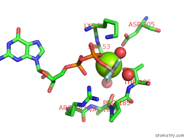
Mono view
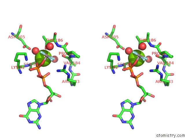
Stereo pair view
Mono view
Stereo pair view
A full contact list of Magnesium with other atoms in the Mg binding
site number 1 of Crystal Structure of Activated G Alpha Q Bound to Its Effector Phospholipase C Beta 3 within 5.0Å range:
|
Reference:
G.L.Waldo,
T.K.Ricks,
S.N.Hicks,
M.L.Cheever,
T.Kawano,
K.Tsuboi,
X.Wang,
C.Montell,
T.Kozasa,
J.Sondek,
T.K.Harden.
Kinetic Scaffolding Mediated By A Phospholipase C-{Beta} and Gq Signaling Complex. Science V. 330 974 2010.
ISSN: ISSN 0036-8075
PubMed: 20966218
DOI: 10.1126/SCIENCE.1193438
Page generated: Mon Dec 14 08:35:05 2020
ISSN: ISSN 0036-8075
PubMed: 20966218
DOI: 10.1126/SCIENCE.1193438
Last articles
Zn in 8WB0Zn in 8WAX
Zn in 8WAU
Zn in 8WAZ
Zn in 8WAY
Zn in 8WAV
Zn in 8WAW
Zn in 8WAT
Zn in 8W7M
Zn in 8WD3