Magnesium in PDB 5iun: Crystal Structure of the Desk-Desr Complex in the Phosphatase State
Enzymatic activity of Crystal Structure of the Desk-Desr Complex in the Phosphatase State
All present enzymatic activity of Crystal Structure of the Desk-Desr Complex in the Phosphatase State:
2.7.13.3;
2.7.13.3;
Protein crystallography data
The structure of Crystal Structure of the Desk-Desr Complex in the Phosphatase State, PDB code: 5iun
was solved by
F.Trajtenberg,
J.A.Imelio,
N.Larrieux,
A.Buschiazzo,
with X-Ray Crystallography technique. A brief refinement statistics is given in the table below:
| Resolution Low / High (Å) | 48.35 / 2.79 |
| Space group | P 31 2 1 |
| Cell size a, b, c (Å), α, β, γ (°) | 94.330, 94.330, 239.910, 90.00, 90.00, 120.00 |
| R / Rfree (%) | 21.3 / 24.8 |
Other elements in 5iun:
The structure of Crystal Structure of the Desk-Desr Complex in the Phosphatase State also contains other interesting chemical elements:
| Fluorine | (F) | 9 atoms |
Magnesium Binding Sites:
The binding sites of Magnesium atom in the Crystal Structure of the Desk-Desr Complex in the Phosphatase State
(pdb code 5iun). This binding sites where shown within
5.0 Angstroms radius around Magnesium atom.
In total 6 binding sites of Magnesium where determined in the Crystal Structure of the Desk-Desr Complex in the Phosphatase State, PDB code: 5iun:
Jump to Magnesium binding site number: 1; 2; 3; 4; 5; 6;
In total 6 binding sites of Magnesium where determined in the Crystal Structure of the Desk-Desr Complex in the Phosphatase State, PDB code: 5iun:
Jump to Magnesium binding site number: 1; 2; 3; 4; 5; 6;
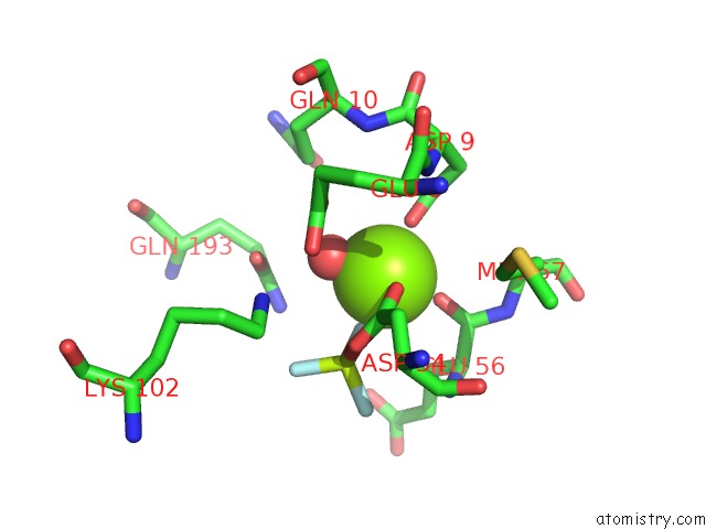
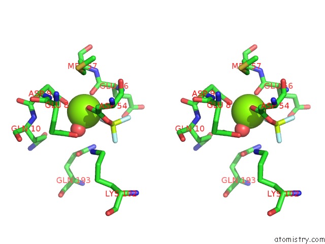
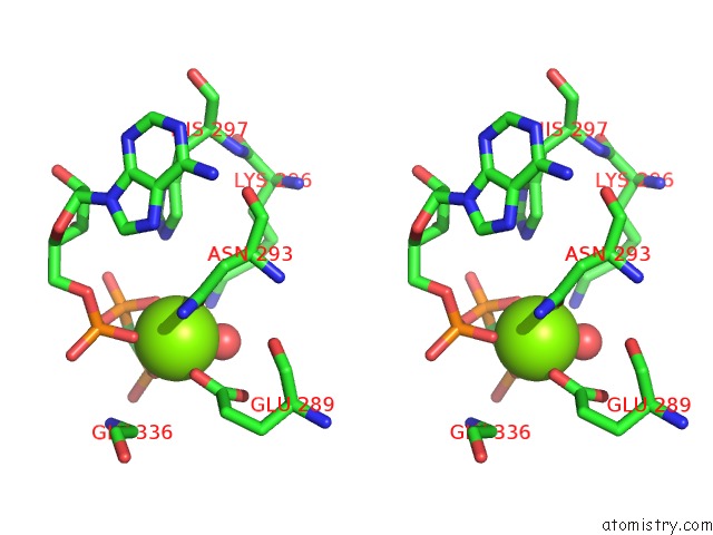
Magnesium binding site 1 out of 6 in 5iun
Go back to
Magnesium binding site 1 out
of 6 in the Crystal Structure of the Desk-Desr Complex in the Phosphatase State
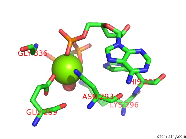
Mono view
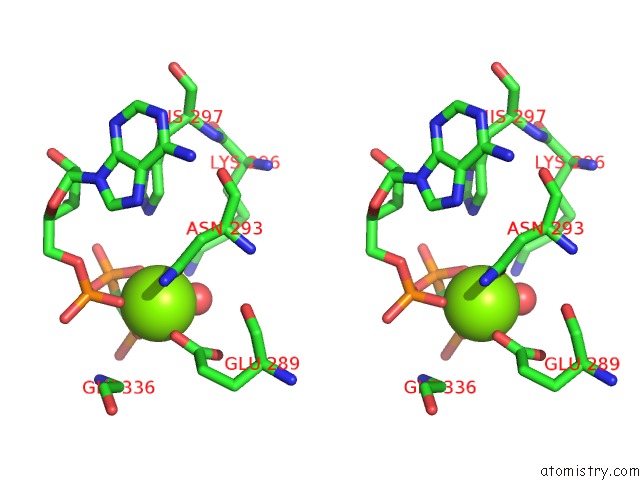
Stereo pair view
Mono view
Stereo pair view
A full contact list of Magnesium with other atoms in the Mg binding
site number 1 of Crystal Structure of the Desk-Desr Complex in the Phosphatase State within 5.0Å range:
|
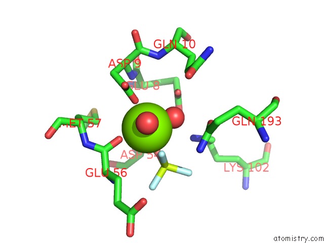
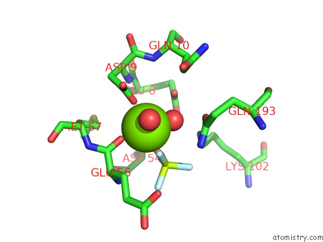
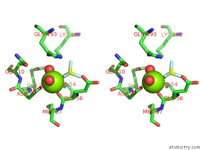
Magnesium binding site 2 out of 6 in 5iun
Go back to
Magnesium binding site 2 out
of 6 in the Crystal Structure of the Desk-Desr Complex in the Phosphatase State
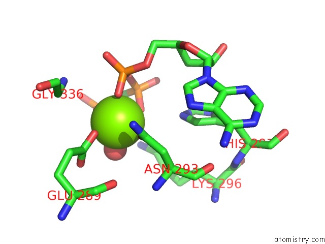
Mono view
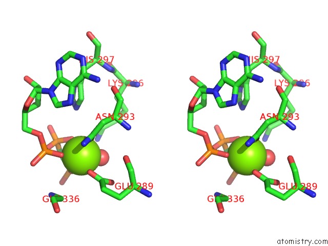
Stereo pair view
Mono view
Stereo pair view
A full contact list of Magnesium with other atoms in the Mg binding
site number 2 of Crystal Structure of the Desk-Desr Complex in the Phosphatase State within 5.0Å range:
|
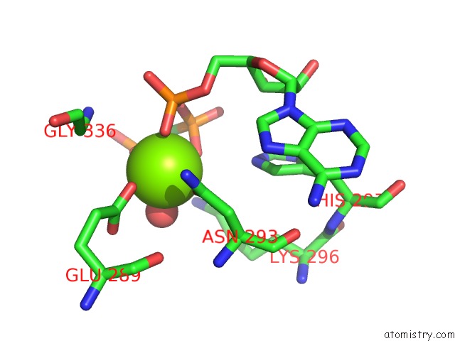
Magnesium binding site 3 out of 6 in 5iun
Go back to
Magnesium binding site 3 out
of 6 in the Crystal Structure of the Desk-Desr Complex in the Phosphatase State
Mono view
Stereo pair view
Mono view
Stereo pair view
A full contact list of Magnesium with other atoms in the Mg binding
site number 3 of Crystal Structure of the Desk-Desr Complex in the Phosphatase State within 5.0Å range:
|
Magnesium binding site 4 out of 6 in 5iun
Go back to
Magnesium binding site 4 out
of 6 in the Crystal Structure of the Desk-Desr Complex in the Phosphatase State
Mono view

Stereo pair view
Mono view
Stereo pair view
A full contact list of Magnesium with other atoms in the Mg binding
site number 4 of Crystal Structure of the Desk-Desr Complex in the Phosphatase State within 5.0Å range:
|
Magnesium binding site 5 out of 6 in 5iun
Go back to
Magnesium binding site 5 out
of 6 in the Crystal Structure of the Desk-Desr Complex in the Phosphatase State
Mono view
Stereo pair view
Mono view
Stereo pair view
A full contact list of Magnesium with other atoms in the Mg binding
site number 5 of Crystal Structure of the Desk-Desr Complex in the Phosphatase State within 5.0Å range:
|
Magnesium binding site 6 out of 6 in 5iun
Go back to
Magnesium binding site 6 out
of 6 in the Crystal Structure of the Desk-Desr Complex in the Phosphatase State
Mono view
Stereo pair view
Mono view
Stereo pair view
A full contact list of Magnesium with other atoms in the Mg binding
site number 6 of Crystal Structure of the Desk-Desr Complex in the Phosphatase State within 5.0Å range:
|
Reference:
F.Trajtenberg,
J.A.Imelio,
M.R.Machado,
N.Larrieux,
M.A.Marti,
G.Obal,
A.E.Mechaly,
A.Buschiazzo.
Regulation of Signaling Directionality Revealed By 3D Snapshots of A Kinase:Regulator Complex in Action. Elife V. 5 2016.
ISSN: ESSN 2050-084X
PubMed: 27938660
DOI: 10.7554/ELIFE.21422
Page generated: Mon Dec 14 20:31:15 2020
ISSN: ESSN 2050-084X
PubMed: 27938660
DOI: 10.7554/ELIFE.21422
Last articles
Zn in 8WB0Zn in 8WAX
Zn in 8WAU
Zn in 8WAZ
Zn in 8WAY
Zn in 8WAV
Zn in 8WAW
Zn in 8WAT
Zn in 8W7M
Zn in 8WD3