Magnesium in PDB 6kpv: Crystal Structures of Na+,K+-Atpase in Complex with Bufalin
Protein crystallography data
The structure of Crystal Structures of Na+,K+-Atpase in Complex with Bufalin, PDB code: 6kpv
was solved by
H.Ogawa,
K.Motoyama,
F.Cornelius,
B.Vilsen,
C.Toyoshima,
with X-Ray Crystallography technique. A brief refinement statistics is given in the table below:
| Resolution Low / High (Å) | 16.00 / 3.72 |
| Space group | P 21 21 21 |
| Cell size a, b, c (Å), α, β, γ (°) | 115.792, 118.032, 495.452, 90.00, 90.00, 90.00 |
| R / Rfree (%) | 23.8 / 28.6 |
Other elements in 6kpv:
The structure of Crystal Structures of Na+,K+-Atpase in Complex with Bufalin also contains other interesting chemical elements:
| Sodium | (Na) | 2 atoms |
Magnesium Binding Sites:
The binding sites of Magnesium atom in the Crystal Structures of Na+,K+-Atpase in Complex with Bufalin
(pdb code 6kpv). This binding sites where shown within
5.0 Angstroms radius around Magnesium atom.
In total 4 binding sites of Magnesium where determined in the Crystal Structures of Na+,K+-Atpase in Complex with Bufalin, PDB code: 6kpv:
Jump to Magnesium binding site number: 1; 2; 3; 4;
In total 4 binding sites of Magnesium where determined in the Crystal Structures of Na+,K+-Atpase in Complex with Bufalin, PDB code: 6kpv:
Jump to Magnesium binding site number: 1; 2; 3; 4;
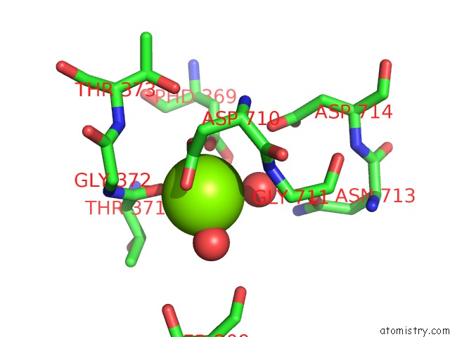
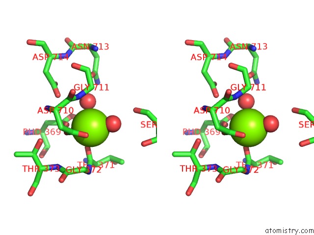
Magnesium binding site 1 out of 4 in 6kpv
Go back to
Magnesium binding site 1 out
of 4 in the Crystal Structures of Na+,K+-Atpase in Complex with Bufalin
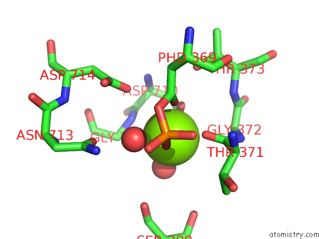
Mono view
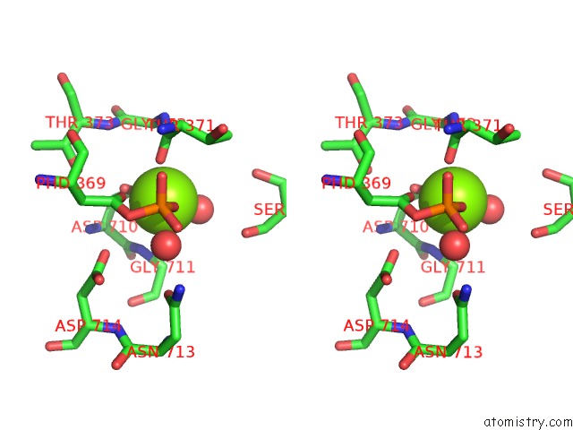
Stereo pair view
Mono view
Stereo pair view
A full contact list of Magnesium with other atoms in the Mg binding
site number 1 of Crystal Structures of Na+,K+-Atpase in Complex with Bufalin within 5.0Å range:
|
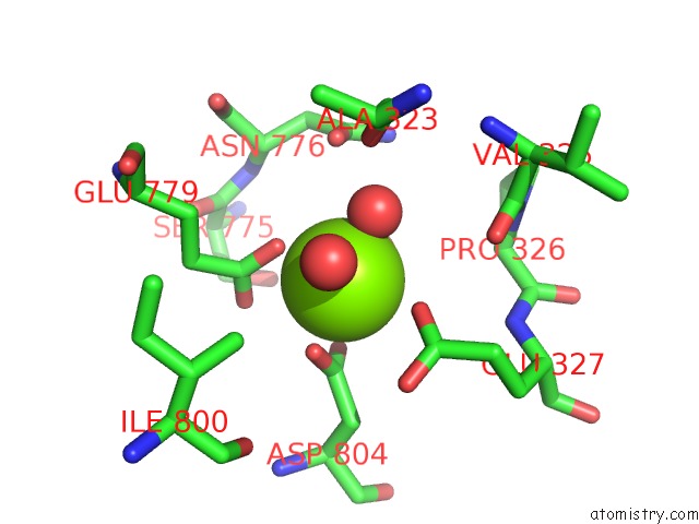
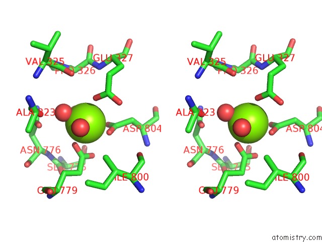
Magnesium binding site 2 out of 4 in 6kpv
Go back to
Magnesium binding site 2 out
of 4 in the Crystal Structures of Na+,K+-Atpase in Complex with Bufalin
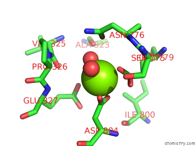
Mono view
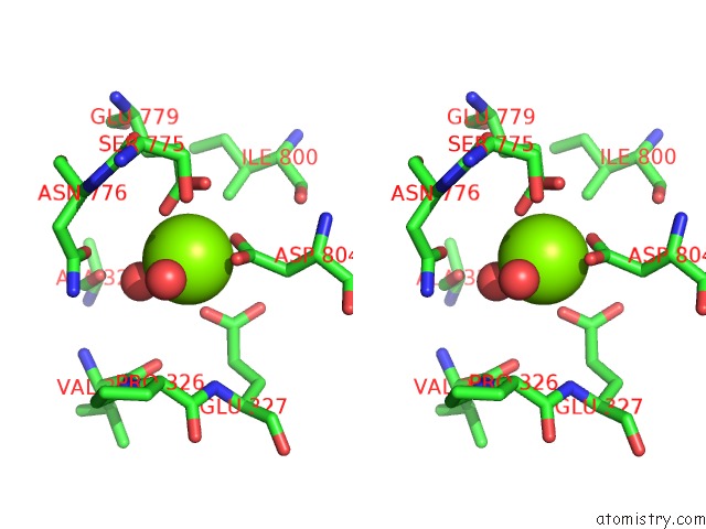
Stereo pair view
Mono view
Stereo pair view
A full contact list of Magnesium with other atoms in the Mg binding
site number 2 of Crystal Structures of Na+,K+-Atpase in Complex with Bufalin within 5.0Å range:
|
Magnesium binding site 3 out of 4 in 6kpv
Go back to
Magnesium binding site 3 out
of 4 in the Crystal Structures of Na+,K+-Atpase in Complex with Bufalin
Mono view
Stereo pair view
Mono view
Stereo pair view
A full contact list of Magnesium with other atoms in the Mg binding
site number 3 of Crystal Structures of Na+,K+-Atpase in Complex with Bufalin within 5.0Å range:
|
Magnesium binding site 4 out of 4 in 6kpv
Go back to
Magnesium binding site 4 out
of 4 in the Crystal Structures of Na+,K+-Atpase in Complex with Bufalin
Mono view
Stereo pair view
Mono view
Stereo pair view
A full contact list of Magnesium with other atoms in the Mg binding
site number 4 of Crystal Structures of Na+,K+-Atpase in Complex with Bufalin within 5.0Å range:
|
Reference:
H.Ogawa,
K.Motoyama,
F.Cornelius,
B.Vilsen,
C.Toyoshima.
X-Ray Crystal Structures of Na+,K+-Atpase in Complex with Cardiotonic Steroids To Be Published.
Page generated: Mon Jan 25 08:48:46 2021
Last articles
Zn in 8WB0Zn in 8WAX
Zn in 8WAU
Zn in 8WAZ
Zn in 8WAY
Zn in 8WAV
Zn in 8WAW
Zn in 8WAT
Zn in 8W7M
Zn in 8WD3