Magnesium in PDB 7n37: Crystal Structure of 3-Site Deamidated Variant of Human Gamma(S)- Crystallin
Protein crystallography data
The structure of Crystal Structure of 3-Site Deamidated Variant of Human Gamma(S)- Crystallin, PDB code: 7n37
was solved by
B.Norton-Baker,
P.Mehrabi,
R.W.Martin,
with X-Ray Crystallography technique. A brief refinement statistics is given in the table below:
| Resolution Low / High (Å) | 27.62 / 1.30 |
| Space group | P 21 21 21 |
| Cell size a, b, c (Å), α, β, γ (°) | 29.449, 62.552, 79.626, 90, 90, 90 |
| R / Rfree (%) | 14.5 / 17.9 |
Magnesium Binding Sites:
The binding sites of Magnesium atom in the Crystal Structure of 3-Site Deamidated Variant of Human Gamma(S)- Crystallin
(pdb code 7n37). This binding sites where shown within
5.0 Angstroms radius around Magnesium atom.
In total only one binding site of Magnesium was determined in the Crystal Structure of 3-Site Deamidated Variant of Human Gamma(S)- Crystallin, PDB code: 7n37:
In total only one binding site of Magnesium was determined in the Crystal Structure of 3-Site Deamidated Variant of Human Gamma(S)- Crystallin, PDB code: 7n37:
Magnesium binding site 1 out of 1 in 7n37
Go back to
Magnesium binding site 1 out
of 1 in the Crystal Structure of 3-Site Deamidated Variant of Human Gamma(S)- Crystallin
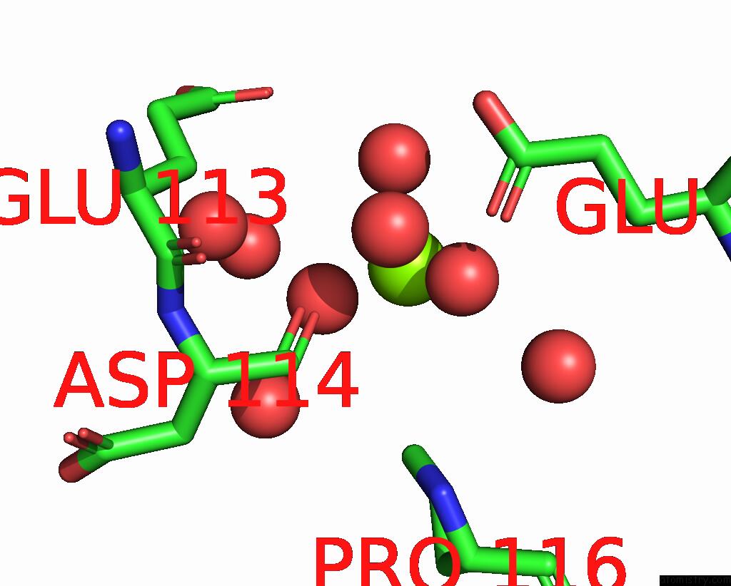
Mono view
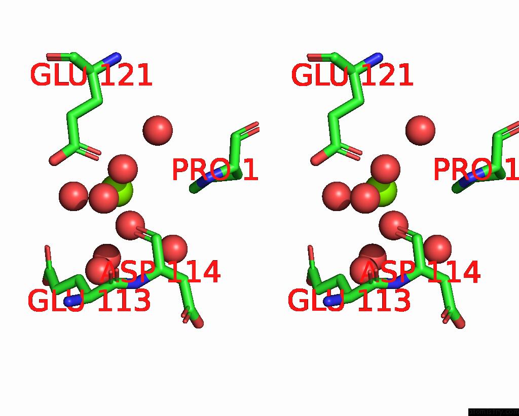
Stereo pair view
Mono view
Stereo pair view
A full contact list of Magnesium with other atoms in the Mg binding
site number 1 of Crystal Structure of 3-Site Deamidated Variant of Human Gamma(S)- Crystallin within 5.0Å range:
|
Reference:
B.Norton-Baker,
P.Mehrabi,
A.O.Kwok,
K.W.Roskamp,
M.A.Rocha,
M.A.Sprague-Piercy,
D.Von Stetten,
R.J.D.Miller,
R.W.Martin.
Deamidation of the Human Eye Lens Protein Gamma S-Crystallin Accelerates Oxidative Aging. Structure V. 30 763 2022.
ISSN: ISSN 0969-2126
PubMed: 35338852
DOI: 10.1016/J.STR.2022.03.002
Page generated: Thu Oct 3 01:13:05 2024
ISSN: ISSN 0969-2126
PubMed: 35338852
DOI: 10.1016/J.STR.2022.03.002
Last articles
Zn in 9J0NZn in 9J0O
Zn in 9J0P
Zn in 9FJX
Zn in 9EKB
Zn in 9C0F
Zn in 9CAH
Zn in 9CH0
Zn in 9CH3
Zn in 9CH1