Magnesium in PDB 7ou4: The Structure of Muts Bound to One Molecule of Atp and One Molecule of Adp
Magnesium Binding Sites:
The binding sites of Magnesium atom in the The Structure of Muts Bound to One Molecule of Atp and One Molecule of Adp
(pdb code 7ou4). This binding sites where shown within
5.0 Angstroms radius around Magnesium atom.
In total 2 binding sites of Magnesium where determined in the The Structure of Muts Bound to One Molecule of Atp and One Molecule of Adp, PDB code: 7ou4:
Jump to Magnesium binding site number: 1; 2;
In total 2 binding sites of Magnesium where determined in the The Structure of Muts Bound to One Molecule of Atp and One Molecule of Adp, PDB code: 7ou4:
Jump to Magnesium binding site number: 1; 2;
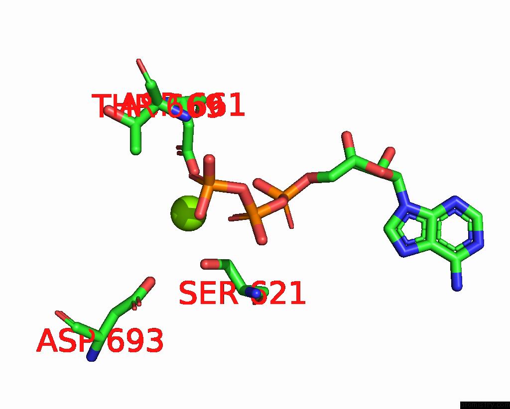
Magnesium binding site 1 out of 2 in 7ou4
Go back to
Magnesium binding site 1 out
of 2 in the The Structure of Muts Bound to One Molecule of Atp and One Molecule of Adp
Mono view

Stereo pair view
Mono view
Stereo pair view
A full contact list of Magnesium with other atoms in the Mg binding
site number 1 of The Structure of Muts Bound to One Molecule of Atp and One Molecule of Adp within 5.0Å range:
|
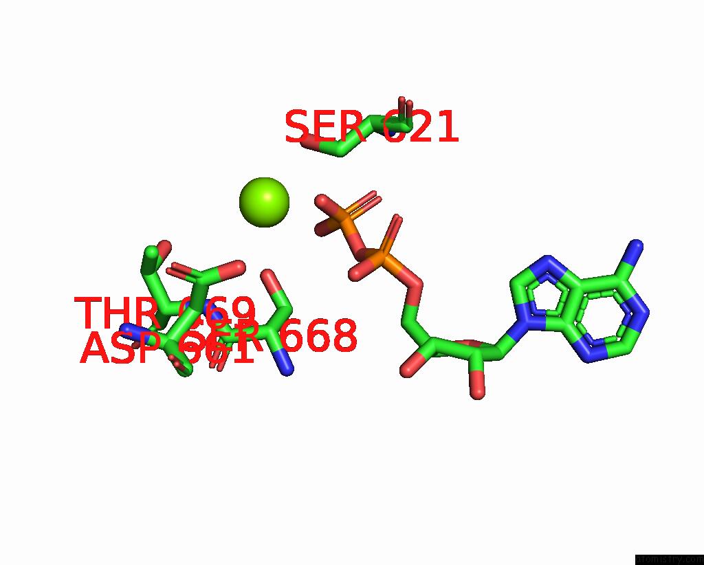
Magnesium binding site 2 out of 2 in 7ou4
Go back to
Magnesium binding site 2 out
of 2 in the The Structure of Muts Bound to One Molecule of Atp and One Molecule of Adp
Mono view

Stereo pair view
Mono view
Stereo pair view
A full contact list of Magnesium with other atoms in the Mg binding
site number 2 of The Structure of Muts Bound to One Molecule of Atp and One Molecule of Adp within 5.0Å range:
|
Reference:
A.Borsellini,
V.Kunetsky,
P.Friedhoff,
M.H.Lamers.
Cryogenic Electron Microscopy Structures Reveal How Atp and Dna Binding in Muts Coordinates Sequential Steps of Dna Mismatch Repair. Nat.Struct.Mol.Biol. V. 29 59 2022.
ISSN: ESSN 1545-9985
PubMed: 35013597
DOI: 10.1038/S41594-021-00707-1
Page generated: Thu Oct 3 03:35:16 2024
ISSN: ESSN 1545-9985
PubMed: 35013597
DOI: 10.1038/S41594-021-00707-1
Last articles
Zn in 9JYWZn in 9IR4
Zn in 9IR3
Zn in 9GMX
Zn in 9GMW
Zn in 9JEJ
Zn in 9ERF
Zn in 9ERE
Zn in 9EGV
Zn in 9EGW