Magnesium in PDB 7pji: Crystal Structure of Pseudomonas Aeruginosa Guab (Imp Dehydrogenase) Bound to Atp and Gdp at 1.65A Resolution
Enzymatic activity of Crystal Structure of Pseudomonas Aeruginosa Guab (Imp Dehydrogenase) Bound to Atp and Gdp at 1.65A Resolution
All present enzymatic activity of Crystal Structure of Pseudomonas Aeruginosa Guab (Imp Dehydrogenase) Bound to Atp and Gdp at 1.65A Resolution:
1.1.1.205;
1.1.1.205;
Protein crystallography data
The structure of Crystal Structure of Pseudomonas Aeruginosa Guab (Imp Dehydrogenase) Bound to Atp and Gdp at 1.65A Resolution, PDB code: 7pji
was solved by
D.Fernandez-Justel,
R.M.Buey,
with X-Ray Crystallography technique. A brief refinement statistics is given in the table below:
| Resolution Low / High (Å) | 50.70 / 1.65 |
| Space group | I 4 |
| Cell size a, b, c (Å), α, β, γ (°) | 120.947, 120.947, 145.487, 90, 90, 90 |
| R / Rfree (%) | 17.2 / 19.5 |
Other elements in 7pji:
The structure of Crystal Structure of Pseudomonas Aeruginosa Guab (Imp Dehydrogenase) Bound to Atp and Gdp at 1.65A Resolution also contains other interesting chemical elements:
| Potassium | (K) | 2 atoms |
Magnesium Binding Sites:
The binding sites of Magnesium atom in the Crystal Structure of Pseudomonas Aeruginosa Guab (Imp Dehydrogenase) Bound to Atp and Gdp at 1.65A Resolution
(pdb code 7pji). This binding sites where shown within
5.0 Angstroms radius around Magnesium atom.
In total 2 binding sites of Magnesium where determined in the Crystal Structure of Pseudomonas Aeruginosa Guab (Imp Dehydrogenase) Bound to Atp and Gdp at 1.65A Resolution, PDB code: 7pji:
Jump to Magnesium binding site number: 1; 2;
In total 2 binding sites of Magnesium where determined in the Crystal Structure of Pseudomonas Aeruginosa Guab (Imp Dehydrogenase) Bound to Atp and Gdp at 1.65A Resolution, PDB code: 7pji:
Jump to Magnesium binding site number: 1; 2;
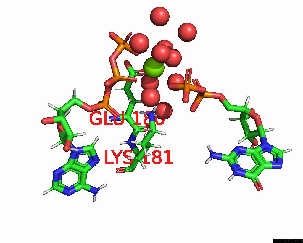
Magnesium binding site 1 out of 2 in 7pji
Go back to
Magnesium binding site 1 out
of 2 in the Crystal Structure of Pseudomonas Aeruginosa Guab (Imp Dehydrogenase) Bound to Atp and Gdp at 1.65A Resolution

Mono view
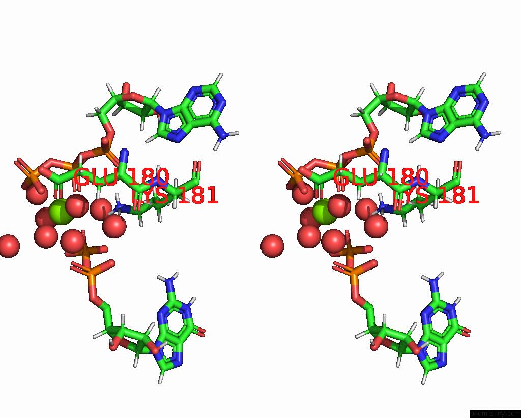
Stereo pair view
Mono view
Stereo pair view
A full contact list of Magnesium with other atoms in the Mg binding
site number 1 of Crystal Structure of Pseudomonas Aeruginosa Guab (Imp Dehydrogenase) Bound to Atp and Gdp at 1.65A Resolution within 5.0Å range:
|
Magnesium binding site 2 out of 2 in 7pji
Go back to
Magnesium binding site 2 out
of 2 in the Crystal Structure of Pseudomonas Aeruginosa Guab (Imp Dehydrogenase) Bound to Atp and Gdp at 1.65A Resolution
Mono view
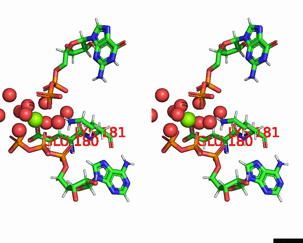
Stereo pair view
Mono view
Stereo pair view
A full contact list of Magnesium with other atoms in the Mg binding
site number 2 of Crystal Structure of Pseudomonas Aeruginosa Guab (Imp Dehydrogenase) Bound to Atp and Gdp at 1.65A Resolution within 5.0Å range:
|
Reference:
D.Fernandez-Justel,
I.Marcos-Alcalde,
F.Abascal,
N.Vidana,
P.Gomez-Puertas,
A.Jimenez,
J.L.Revuelta,
R.M.Buey.
Diversity of Mechanisms to Control Bacterial Gtp Homeostasis By the Mutually Exclusive Binding of Adenine and Guanine Nucleotides to Imp Dehydrogenase. Protein Sci. V. 31 E4314 2022.
ISSN: ESSN 1469-896X
PubMed: 35481629
DOI: 10.1002/PRO.4314
Page generated: Thu Oct 3 04:10:46 2024
ISSN: ESSN 1469-896X
PubMed: 35481629
DOI: 10.1002/PRO.4314
Last articles
Zn in 9JYWZn in 9IR4
Zn in 9IR3
Zn in 9GMX
Zn in 9GMW
Zn in 9JEJ
Zn in 9ERF
Zn in 9ERE
Zn in 9EGV
Zn in 9EGW