Magnesium in PDB 7qpi: Structure of Lamprey Vdr in Complex with 1,25D3
Enzymatic activity of Structure of Lamprey Vdr in Complex with 1,25D3
All present enzymatic activity of Structure of Lamprey Vdr in Complex with 1,25D3:
2.3.1.48;
2.3.1.48;
Protein crystallography data
The structure of Structure of Lamprey Vdr in Complex with 1,25D3, PDB code: 7qpi
was solved by
N.Rochel,
with X-Ray Crystallography technique. A brief refinement statistics is given in the table below:
| Resolution Low / High (Å) | 24.76 / 2.50 |
| Space group | P 21 21 21 |
| Cell size a, b, c (Å), α, β, γ (°) | 37.32, 66.17, 105.69, 90, 90, 90 |
| R / Rfree (%) | 18.3 / 25.6 |
Magnesium Binding Sites:
The binding sites of Magnesium atom in the Structure of Lamprey Vdr in Complex with 1,25D3
(pdb code 7qpi). This binding sites where shown within
5.0 Angstroms radius around Magnesium atom.
In total 2 binding sites of Magnesium where determined in the Structure of Lamprey Vdr in Complex with 1,25D3, PDB code: 7qpi:
Jump to Magnesium binding site number: 1; 2;
In total 2 binding sites of Magnesium where determined in the Structure of Lamprey Vdr in Complex with 1,25D3, PDB code: 7qpi:
Jump to Magnesium binding site number: 1; 2;
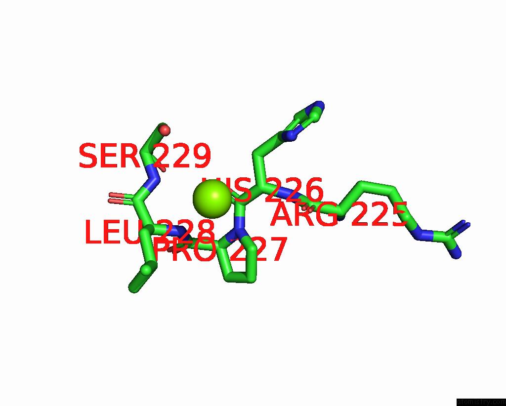
Magnesium binding site 1 out of 2 in 7qpi
Go back to
Magnesium binding site 1 out
of 2 in the Structure of Lamprey Vdr in Complex with 1,25D3
Mono view

Stereo pair view
Mono view
Stereo pair view
A full contact list of Magnesium with other atoms in the Mg binding
site number 1 of Structure of Lamprey Vdr in Complex with 1,25D3 within 5.0Å range:
|
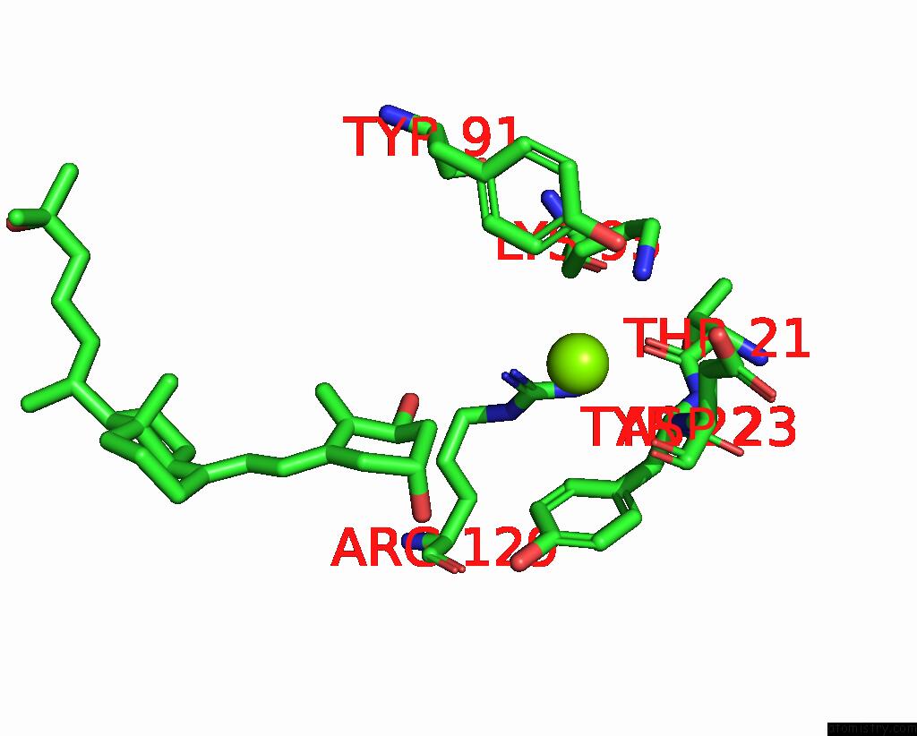
Magnesium binding site 2 out of 2 in 7qpi
Go back to
Magnesium binding site 2 out
of 2 in the Structure of Lamprey Vdr in Complex with 1,25D3
Mono view

Stereo pair view
Mono view
Stereo pair view
A full contact list of Magnesium with other atoms in the Mg binding
site number 2 of Structure of Lamprey Vdr in Complex with 1,25D3 within 5.0Å range:
|
Reference:
R.Sigueiro,
L.Bianchetti,
C.Peluso-Iltis,
S.Chalhoub,
A.Dejaegere,
J.Osz,
N.Rochel.
Advances in Vitamin D Receptor Function and Evolution Based on the 3D Structure of the Lamprey Ligand-Binding Domain. J.Med.Chem. V. 65 5821 2022.
ISSN: ISSN 0022-2623
PubMed: 35302785
DOI: 10.1021/ACS.JMEDCHEM.2C00171
Page generated: Thu Oct 3 05:18:53 2024
ISSN: ISSN 0022-2623
PubMed: 35302785
DOI: 10.1021/ACS.JMEDCHEM.2C00171
Last articles
Zn in 9J0NZn in 9J0O
Zn in 9J0P
Zn in 9FJX
Zn in 9EKB
Zn in 9C0F
Zn in 9CAH
Zn in 9CH0
Zn in 9CH3
Zn in 9CH1