Magnesium in PDB 7u0y: Crystal Structure of Pepper Rna Aptamer in Complex with HBC599 Ligand and Fab BL3-6
Protein crystallography data
The structure of Crystal Structure of Pepper Rna Aptamer in Complex with HBC599 Ligand and Fab BL3-6, PDB code: 7u0y
was solved by
H.C.Rees,
J.A.Piccirilli,
with X-Ray Crystallography technique. A brief refinement statistics is given in the table below:
| Resolution Low / High (Å) | 58.67 / 2.66 |
| Space group | P 21 21 21 |
| Cell size a, b, c (Å), α, β, γ (°) | 61.247, 96.071, 148.165, 90, 90, 90 |
| R / Rfree (%) | 21.9 / 25.5 |
Magnesium Binding Sites:
The binding sites of Magnesium atom in the Crystal Structure of Pepper Rna Aptamer in Complex with HBC599 Ligand and Fab BL3-6
(pdb code 7u0y). This binding sites where shown within
5.0 Angstroms radius around Magnesium atom.
In total 2 binding sites of Magnesium where determined in the Crystal Structure of Pepper Rna Aptamer in Complex with HBC599 Ligand and Fab BL3-6, PDB code: 7u0y:
Jump to Magnesium binding site number: 1; 2;
In total 2 binding sites of Magnesium where determined in the Crystal Structure of Pepper Rna Aptamer in Complex with HBC599 Ligand and Fab BL3-6, PDB code: 7u0y:
Jump to Magnesium binding site number: 1; 2;
Magnesium binding site 1 out of 2 in 7u0y
Go back to
Magnesium binding site 1 out
of 2 in the Crystal Structure of Pepper Rna Aptamer in Complex with HBC599 Ligand and Fab BL3-6
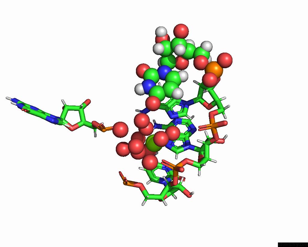
Mono view
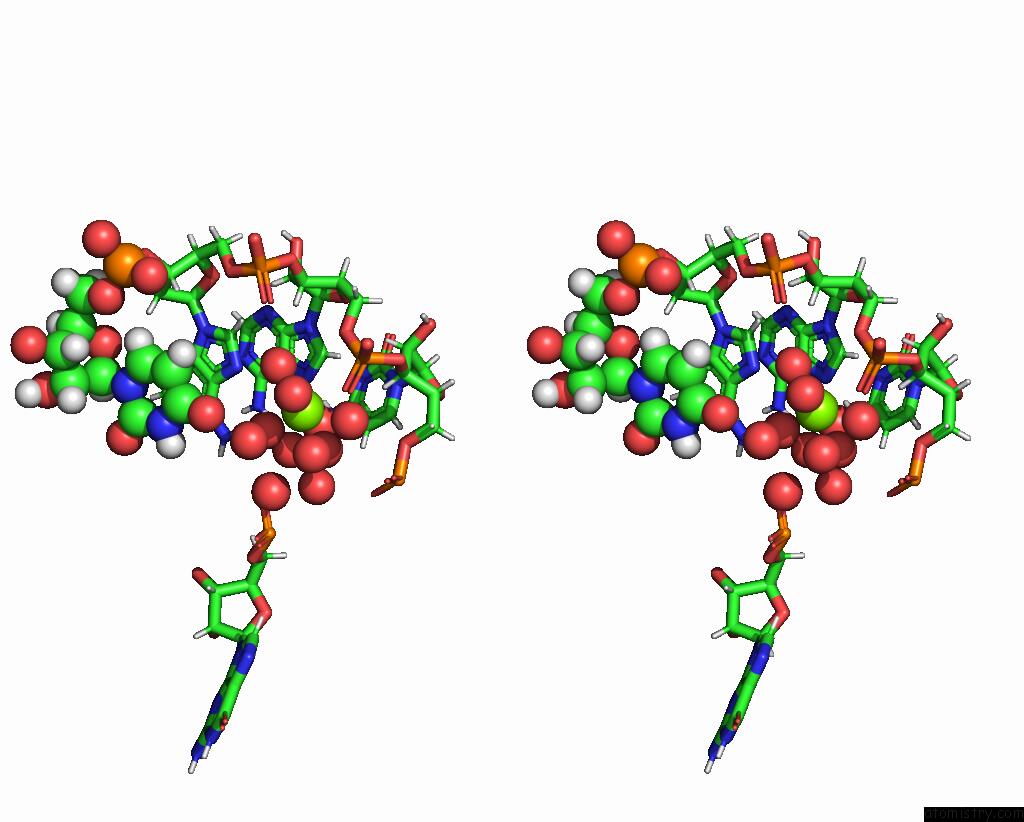
Stereo pair view
Mono view
Stereo pair view
A full contact list of Magnesium with other atoms in the Mg binding
site number 1 of Crystal Structure of Pepper Rna Aptamer in Complex with HBC599 Ligand and Fab BL3-6 within 5.0Å range:
|
Magnesium binding site 2 out of 2 in 7u0y
Go back to
Magnesium binding site 2 out
of 2 in the Crystal Structure of Pepper Rna Aptamer in Complex with HBC599 Ligand and Fab BL3-6

Mono view
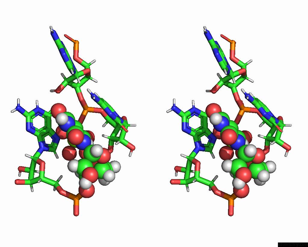
Stereo pair view
Mono view
Stereo pair view
A full contact list of Magnesium with other atoms in the Mg binding
site number 2 of Crystal Structure of Pepper Rna Aptamer in Complex with HBC599 Ligand and Fab BL3-6 within 5.0Å range:
|
Reference:
H.C.Rees,
W.Gogacz,
N.S.Li,
D.Koirala,
J.A.Piccirilli.
Structural Basis For Fluorescence Activation By Pepper Rna. Acs Chem.Biol. V. 17 1866 2022.
ISSN: ESSN 1554-8937
PubMed: 35759696
DOI: 10.1021/ACSCHEMBIO.2C00290
Page generated: Thu Oct 3 09:38:04 2024
ISSN: ESSN 1554-8937
PubMed: 35759696
DOI: 10.1021/ACSCHEMBIO.2C00290
Last articles
Zn in 9J0NZn in 9J0O
Zn in 9J0P
Zn in 9FJX
Zn in 9EKB
Zn in 9C0F
Zn in 9CAH
Zn in 9CH0
Zn in 9CH3
Zn in 9CH1