Magnesium in PDB 7uq1: The X-Ray Crystal Structure of the N-Terminal Domain of Staphylococcus Aureus Fatty Acid Kinase A (Faka, Residues 1-208) in Complex with Amp and A Single Mg Ion at the Dinuclear Binding Site
Protein crystallography data
The structure of The X-Ray Crystal Structure of the N-Terminal Domain of Staphylococcus Aureus Fatty Acid Kinase A (Faka, Residues 1-208) in Complex with Amp and A Single Mg Ion at the Dinuclear Binding Site, PDB code: 7uq1
was solved by
M.G.Cuypers,
C.Subramanian,
J.Seetharaman,
C.O.Rock,
S.W.White,
with X-Ray Crystallography technique. A brief refinement statistics is given in the table below:
| Resolution Low / High (Å) | 29.25 / 1.72 |
| Space group | P 21 21 21 |
| Cell size a, b, c (Å), α, β, γ (°) | 41.843, 54.289, 81.824, 90, 90, 90 |
| R / Rfree (%) | 17.4 / 22.3 |
Magnesium Binding Sites:
The binding sites of Magnesium atom in the The X-Ray Crystal Structure of the N-Terminal Domain of Staphylococcus Aureus Fatty Acid Kinase A (Faka, Residues 1-208) in Complex with Amp and A Single Mg Ion at the Dinuclear Binding Site
(pdb code 7uq1). This binding sites where shown within
5.0 Angstroms radius around Magnesium atom.
In total 3 binding sites of Magnesium where determined in the The X-Ray Crystal Structure of the N-Terminal Domain of Staphylococcus Aureus Fatty Acid Kinase A (Faka, Residues 1-208) in Complex with Amp and A Single Mg Ion at the Dinuclear Binding Site, PDB code: 7uq1:
Jump to Magnesium binding site number: 1; 2; 3;
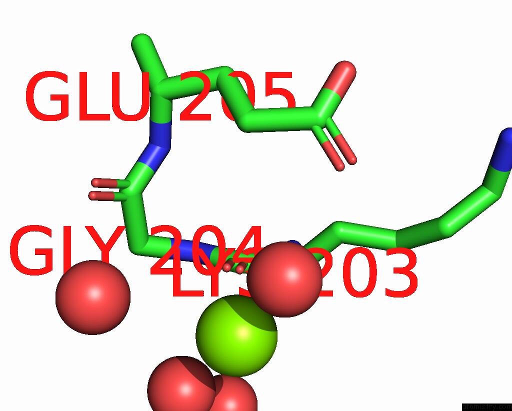
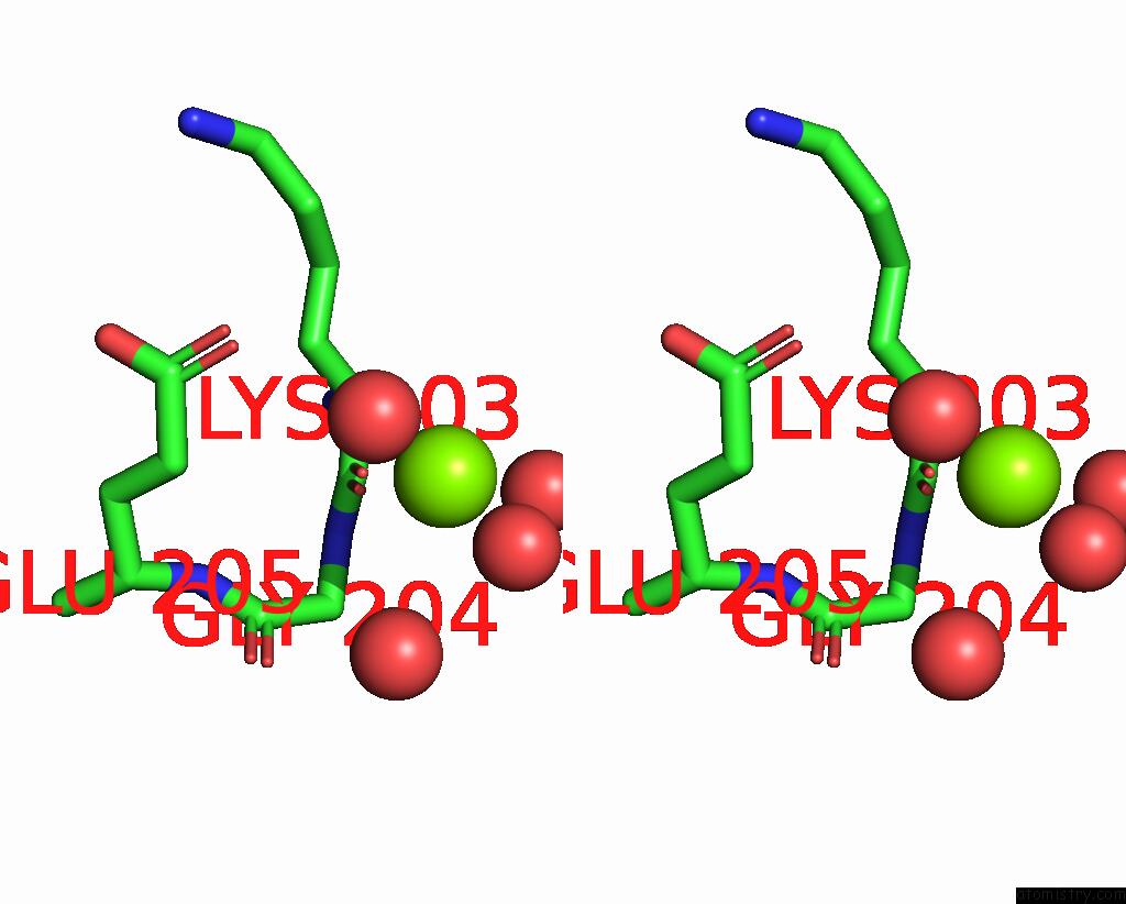
Magnesium binding site 1 out of 3 in 7uq1
Go back to
Magnesium binding site 1 out
of 3 in the The X-Ray Crystal Structure of the N-Terminal Domain of Staphylococcus Aureus Fatty Acid Kinase A (Faka, Residues 1-208) in Complex with Amp and A Single Mg Ion at the Dinuclear Binding Site
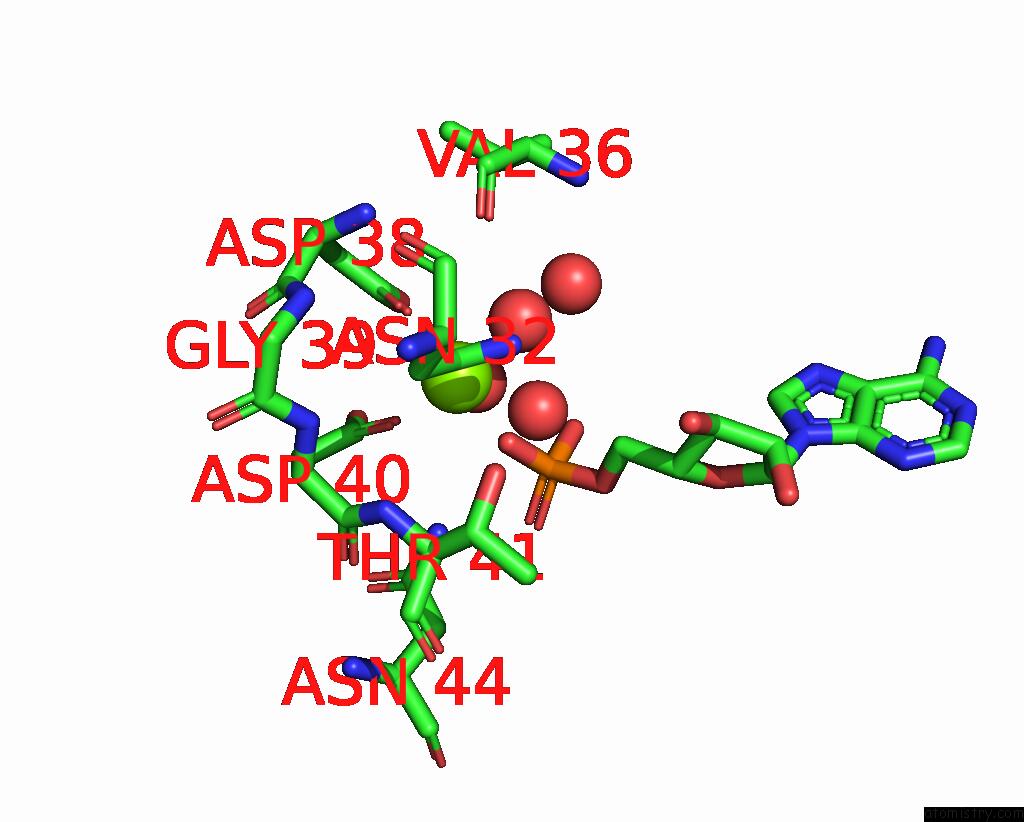
Mono view
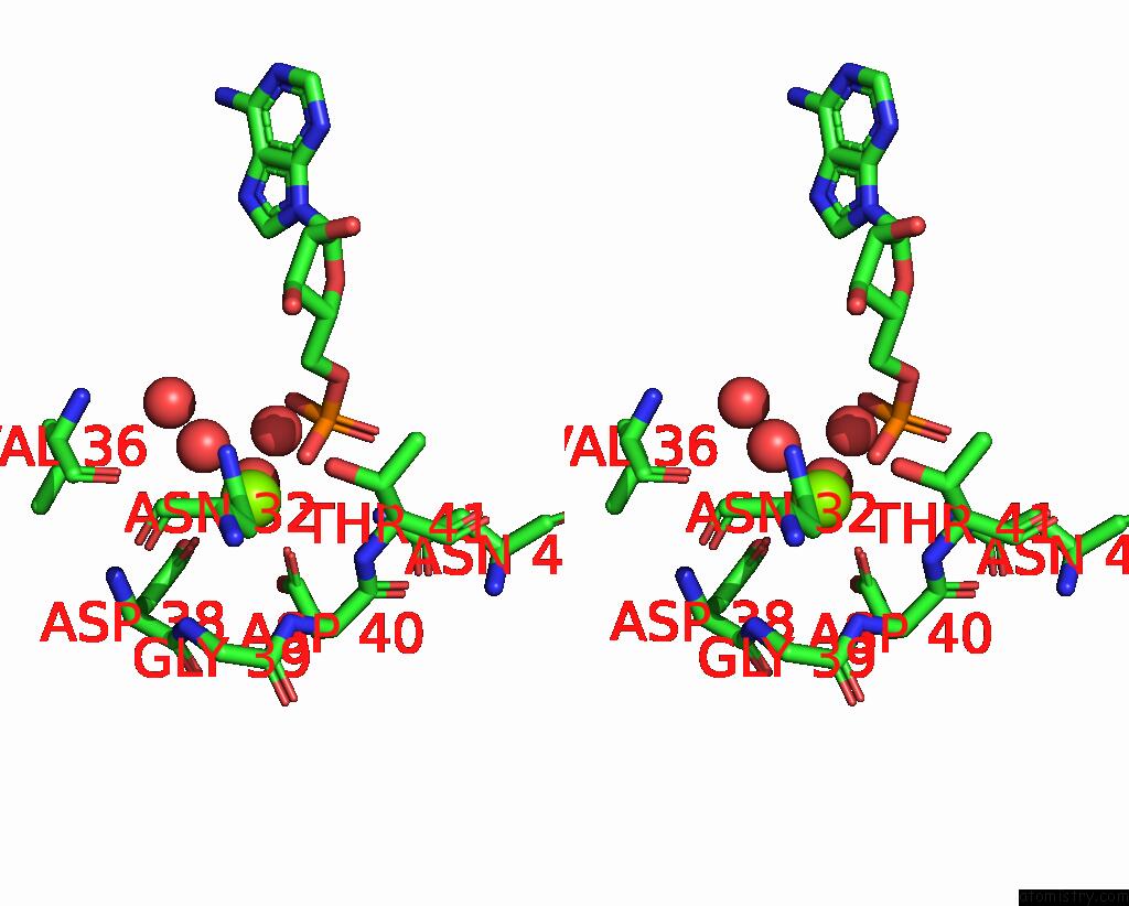
Stereo pair view
Mono view
Stereo pair view
A full contact list of Magnesium with other atoms in the Mg binding
site number 1 of The X-Ray Crystal Structure of the N-Terminal Domain of Staphylococcus Aureus Fatty Acid Kinase A (Faka, Residues 1-208) in Complex with Amp and A Single Mg Ion at the Dinuclear Binding Site within 5.0Å range:
|
Magnesium binding site 2 out of 3 in 7uq1
Go back to
Magnesium binding site 2 out
of 3 in the The X-Ray Crystal Structure of the N-Terminal Domain of Staphylococcus Aureus Fatty Acid Kinase A (Faka, Residues 1-208) in Complex with Amp and A Single Mg Ion at the Dinuclear Binding Site
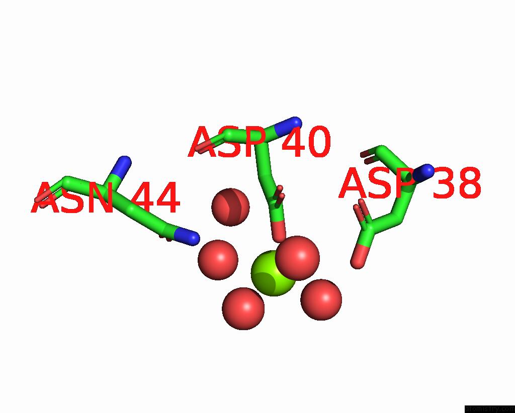
Mono view
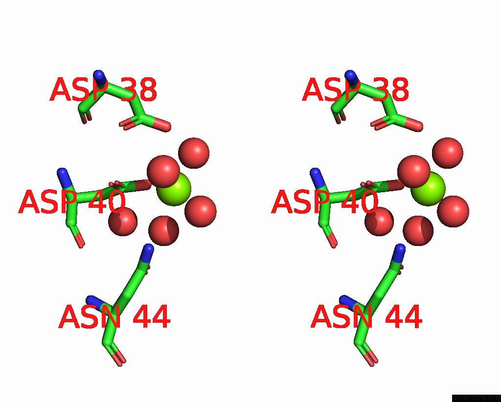
Stereo pair view
Mono view
Stereo pair view
A full contact list of Magnesium with other atoms in the Mg binding
site number 2 of The X-Ray Crystal Structure of the N-Terminal Domain of Staphylococcus Aureus Fatty Acid Kinase A (Faka, Residues 1-208) in Complex with Amp and A Single Mg Ion at the Dinuclear Binding Site within 5.0Å range:
|
Magnesium binding site 3 out of 3 in 7uq1
Go back to
Magnesium binding site 3 out
of 3 in the The X-Ray Crystal Structure of the N-Terminal Domain of Staphylococcus Aureus Fatty Acid Kinase A (Faka, Residues 1-208) in Complex with Amp and A Single Mg Ion at the Dinuclear Binding Site
Mono view
Stereo pair view
Mono view
Stereo pair view
A full contact list of Magnesium with other atoms in the Mg binding
site number 3 of The X-Ray Crystal Structure of the N-Terminal Domain of Staphylococcus Aureus Fatty Acid Kinase A (Faka, Residues 1-208) in Complex with Amp and A Single Mg Ion at the Dinuclear Binding Site within 5.0Å range:
|
Reference:
M.G.Cuypers,
C.Subramanian,
J.Seetharaman,
C.O.Rock,
S.W.White.
The X-Ray Crystal Structure of the N-Terminal Domain of Staphylococcus Aureus Fatty Acid Kinase A (Faka, Residues 1-208) in Complex with Amp and A Single Mg Atom in the Dinuclear Binding Site. To Be Published.
Page generated: Thu Oct 3 10:10:04 2024
Last articles
Zn in 9J0NZn in 9J0O
Zn in 9J0P
Zn in 9FJX
Zn in 9EKB
Zn in 9C0F
Zn in 9CAH
Zn in 9CH0
Zn in 9CH3
Zn in 9CH1