Magnesium in PDB 7w8d: The Structure of Deinococcus Radiodurans Ruvc
Enzymatic activity of The Structure of Deinococcus Radiodurans Ruvc
All present enzymatic activity of The Structure of Deinococcus Radiodurans Ruvc:
3.1.22.4;
3.1.22.4;
Protein crystallography data
The structure of The Structure of Deinococcus Radiodurans Ruvc, PDB code: 7w8d
was solved by
K.Cheng,
with X-Ray Crystallography technique. A brief refinement statistics is given in the table below:
| Resolution Low / High (Å) | 27.71 / 2.75 |
| Space group | P 21 21 21 |
| Cell size a, b, c (Å), α, β, γ (°) | 41.2, 72.77, 112.3, 90, 90, 90 |
| R / Rfree (%) | 27.3 / 30.6 |
Magnesium Binding Sites:
The binding sites of Magnesium atom in the The Structure of Deinococcus Radiodurans Ruvc
(pdb code 7w8d). This binding sites where shown within
5.0 Angstroms radius around Magnesium atom.
In total only one binding site of Magnesium was determined in the The Structure of Deinococcus Radiodurans Ruvc, PDB code: 7w8d:
In total only one binding site of Magnesium was determined in the The Structure of Deinococcus Radiodurans Ruvc, PDB code: 7w8d:
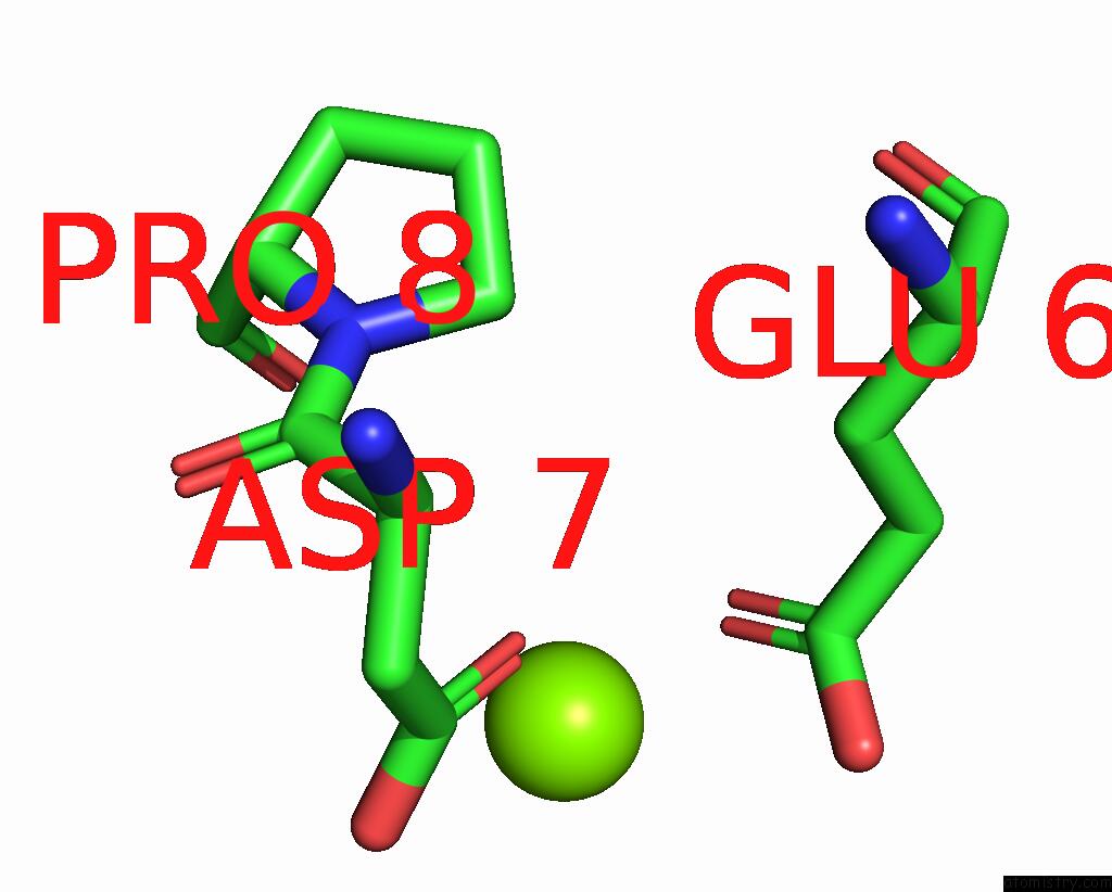
Magnesium binding site 1 out of 1 in 7w8d
Go back to
Magnesium binding site 1 out
of 1 in the The Structure of Deinococcus Radiodurans Ruvc
Mono view

Stereo pair view
Mono view
Stereo pair view
A full contact list of Magnesium with other atoms in the Mg binding
site number 1 of The Structure of Deinococcus Radiodurans Ruvc within 5.0Å range:
|
Reference:
Y.Sun,
J.Yang,
G.Xu,
K.Cheng.
Biochemical and Structural Study of Ruvc and Yqgf From Deinococcus Radiodurans. Mbio V. 13 83422 2022.
ISSN: ESSN 2150-7511
PubMed: 36000732
DOI: 10.1128/MBIO.01834-22
Page generated: Thu Oct 3 11:12:45 2024
ISSN: ESSN 2150-7511
PubMed: 36000732
DOI: 10.1128/MBIO.01834-22
Last articles
K in 9G9VK in 9DTR
K in 9C46
K in 9G9W
K in 9G9X
K in 9ESI
K in 9ESH
K in 8ZEX
K in 8VAV
K in 8VAZ