Magnesium in PDB 7wl6: Crystal Structure of I73L Mutated Human Transthyretin
Protein crystallography data
The structure of Crystal Structure of I73L Mutated Human Transthyretin, PDB code: 7wl6
was solved by
M.Nakagawa,
T.Obita,
M.Mizuguchi,
with X-Ray Crystallography technique. A brief refinement statistics is given in the table below:
| Resolution Low / High (Å) | 35.67 / 1.42 |
| Space group | P 21 21 2 |
| Cell size a, b, c (Å), α, β, γ (°) | 42.84, 85.84, 64.16, 90, 90, 90 |
| R / Rfree (%) | 18.4 / 21 |
Magnesium Binding Sites:
The binding sites of Magnesium atom in the Crystal Structure of I73L Mutated Human Transthyretin
(pdb code 7wl6). This binding sites where shown within
5.0 Angstroms radius around Magnesium atom.
In total 2 binding sites of Magnesium where determined in the Crystal Structure of I73L Mutated Human Transthyretin, PDB code: 7wl6:
Jump to Magnesium binding site number: 1; 2;
In total 2 binding sites of Magnesium where determined in the Crystal Structure of I73L Mutated Human Transthyretin, PDB code: 7wl6:
Jump to Magnesium binding site number: 1; 2;
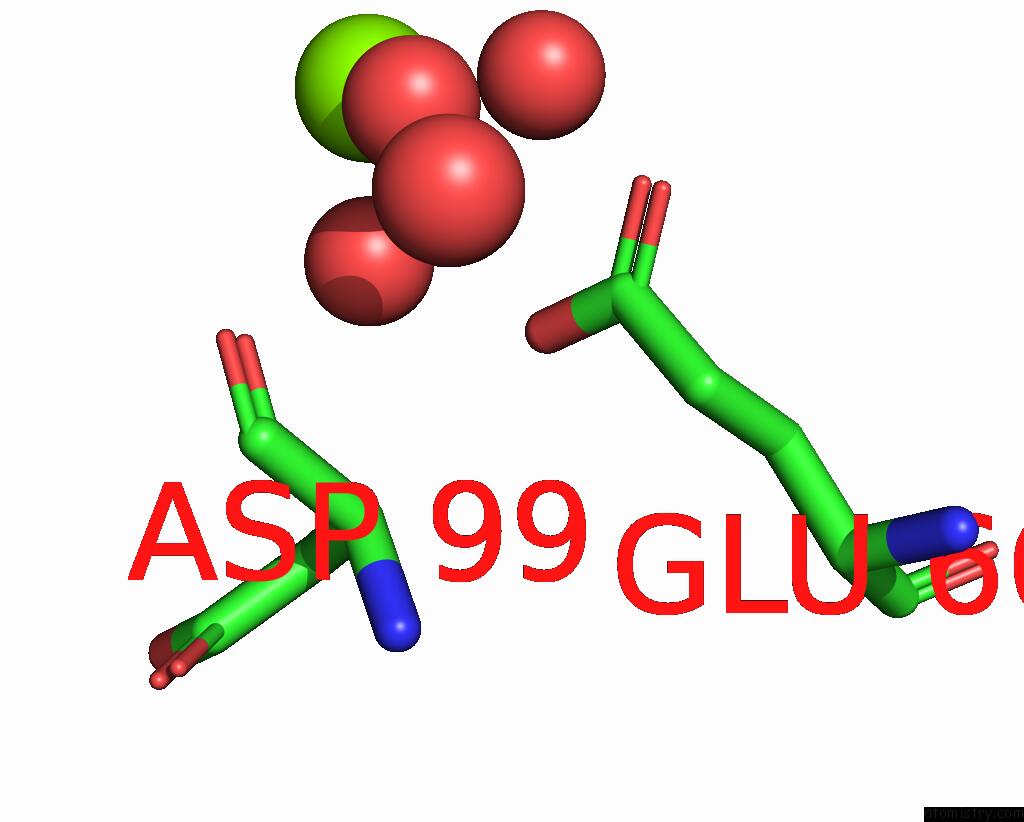
Magnesium binding site 1 out of 2 in 7wl6
Go back to
Magnesium binding site 1 out
of 2 in the Crystal Structure of I73L Mutated Human Transthyretin
Mono view

Stereo pair view
Mono view
Stereo pair view
A full contact list of Magnesium with other atoms in the Mg binding
site number 1 of Crystal Structure of I73L Mutated Human Transthyretin within 5.0Å range:
|
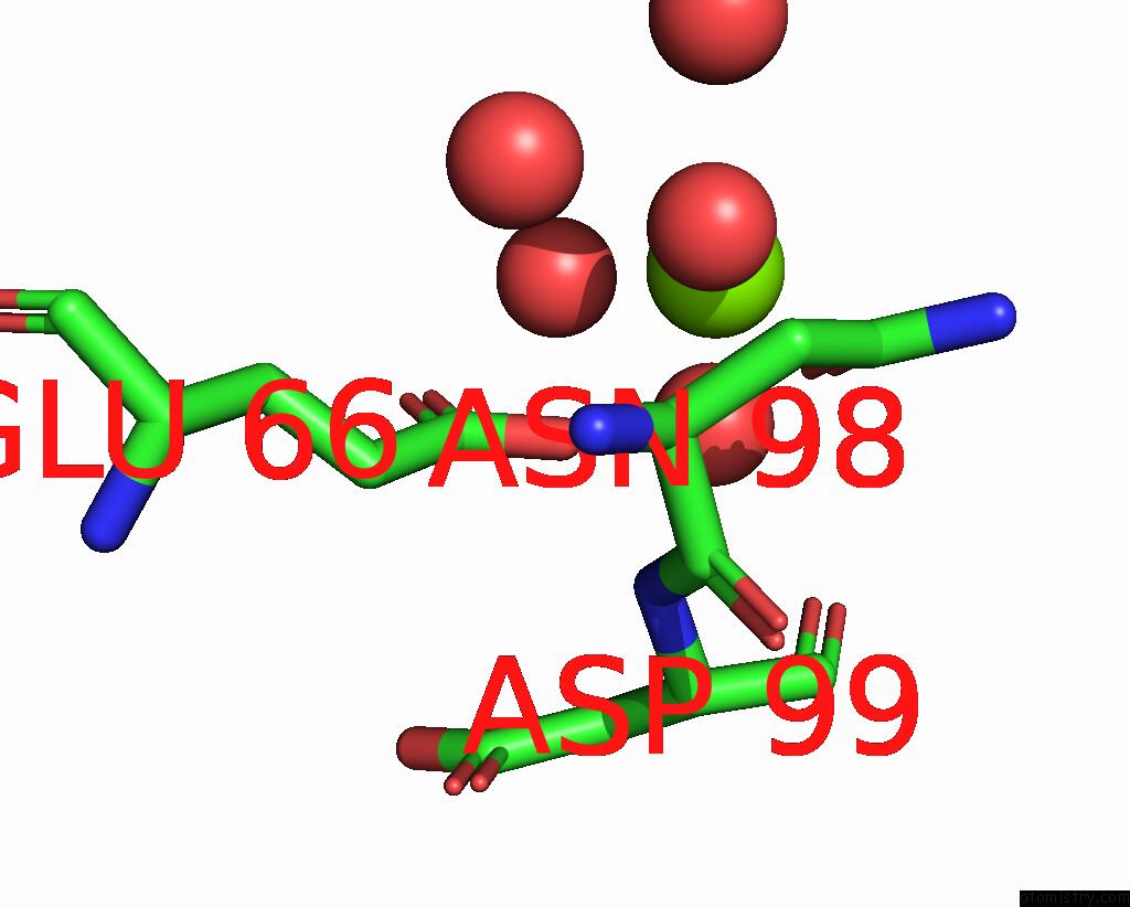
Magnesium binding site 2 out of 2 in 7wl6
Go back to
Magnesium binding site 2 out
of 2 in the Crystal Structure of I73L Mutated Human Transthyretin
Mono view

Stereo pair view
Mono view
Stereo pair view
A full contact list of Magnesium with other atoms in the Mg binding
site number 2 of Crystal Structure of I73L Mutated Human Transthyretin within 5.0Å range:
|
Reference:
M.Nakagawa,
T.Obita,
M.Mizuguchi.
The Hydrophobic Residue LEU73 Is Crucial For the High Stability and Low Aggregation Properties of Murine Transthyretin. Biochem.J. V. 479 1999 2022.
ISSN: ESSN 1470-8728
PubMed: 36098398
DOI: 10.1042/BCJ20220203
Page generated: Thu Oct 3 11:19:58 2024
ISSN: ESSN 1470-8728
PubMed: 36098398
DOI: 10.1042/BCJ20220203
Last articles
Fe in 9EBRFe in 9CU2
Fe in 9CU1
Fe in 9CU0
Fe in 9CJE
Fe in 9CJB
Fe in 9CJF
Fe in 9CTZ
Fe in 9CJD
Fe in 9CJC