Magnesium in PDB 7wyg: Crystal Structure of P450BSBETA-L78I/Q85H/G290I Variant in Complex with Palmitic Acid.
Protein crystallography data
The structure of Crystal Structure of P450BSBETA-L78I/Q85H/G290I Variant in Complex with Palmitic Acid., PDB code: 7wyg
was solved by
F.Li,
C.He,
X.Wang,
with X-Ray Crystallography technique. A brief refinement statistics is given in the table below:
| Resolution Low / High (Å) | 50.98 / 2.00 |
| Space group | P 3 2 1 |
| Cell size a, b, c (Å), α, β, γ (°) | 125.646, 125.646, 145.891, 90, 90, 120 |
| R / Rfree (%) | 17.4 / 20.8 |
Other elements in 7wyg:
The structure of Crystal Structure of P450BSBETA-L78I/Q85H/G290I Variant in Complex with Palmitic Acid. also contains other interesting chemical elements:
| Iron | (Fe) | 2 atoms |
Magnesium Binding Sites:
The binding sites of Magnesium atom in the Crystal Structure of P450BSBETA-L78I/Q85H/G290I Variant in Complex with Palmitic Acid.
(pdb code 7wyg). This binding sites where shown within
5.0 Angstroms radius around Magnesium atom.
In total 2 binding sites of Magnesium where determined in the Crystal Structure of P450BSBETA-L78I/Q85H/G290I Variant in Complex with Palmitic Acid., PDB code: 7wyg:
Jump to Magnesium binding site number: 1; 2;
In total 2 binding sites of Magnesium where determined in the Crystal Structure of P450BSBETA-L78I/Q85H/G290I Variant in Complex with Palmitic Acid., PDB code: 7wyg:
Jump to Magnesium binding site number: 1; 2;
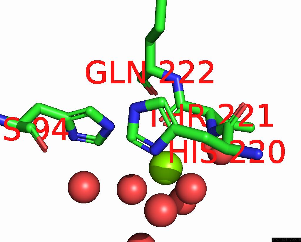
Magnesium binding site 1 out of 2 in 7wyg
Go back to
Magnesium binding site 1 out
of 2 in the Crystal Structure of P450BSBETA-L78I/Q85H/G290I Variant in Complex with Palmitic Acid.

Mono view

Stereo pair view
Mono view
Stereo pair view
A full contact list of Magnesium with other atoms in the Mg binding
site number 1 of Crystal Structure of P450BSBETA-L78I/Q85H/G290I Variant in Complex with Palmitic Acid. within 5.0Å range:
|
Magnesium binding site 2 out of 2 in 7wyg
Go back to
Magnesium binding site 2 out
of 2 in the Crystal Structure of P450BSBETA-L78I/Q85H/G290I Variant in Complex with Palmitic Acid.
Mono view
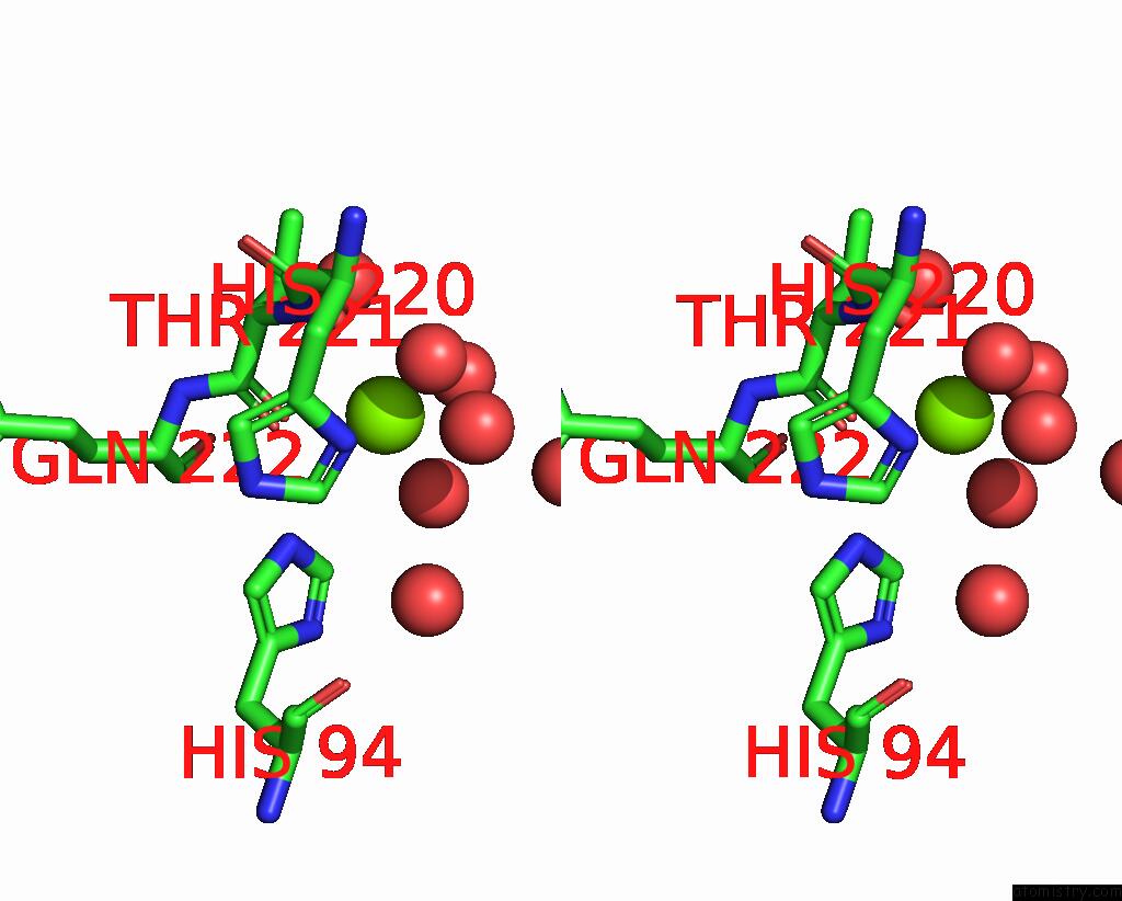
Stereo pair view
Mono view
Stereo pair view
A full contact list of Magnesium with other atoms in the Mg binding
site number 2 of Crystal Structure of P450BSBETA-L78I/Q85H/G290I Variant in Complex with Palmitic Acid. within 5.0Å range:
|
Reference:
K.Zhang,
A.Yu,
X.Chu,
F.Li,
J.Liu,
L.Liu,
W.J.Bai,
C.He,
X.Wang.
Biocatalytic Enantioselective Beta-Hydroxylation of Unactivated C-H Bonds in Aliphatic Carboxylic Acids. Angew.Chem.Int.Ed.Engl. V. 61 04290 2022.
ISSN: ESSN 1521-3773
PubMed: 35536725
DOI: 10.1002/ANIE.202204290
Page generated: Thu Oct 3 11:22:57 2024
ISSN: ESSN 1521-3773
PubMed: 35536725
DOI: 10.1002/ANIE.202204290
Last articles
Zn in 9J0NZn in 9J0O
Zn in 9J0P
Zn in 9FJX
Zn in 9EKB
Zn in 9C0F
Zn in 9CAH
Zn in 9CH0
Zn in 9CH3
Zn in 9CH1