Magnesium in PDB 7yke: Crystal Structure of Chondroitin Abc Lyase I in Complex with Chondroitin Disaccharide 4,6-Sulfate
Enzymatic activity of Crystal Structure of Chondroitin Abc Lyase I in Complex with Chondroitin Disaccharide 4,6-Sulfate
All present enzymatic activity of Crystal Structure of Chondroitin Abc Lyase I in Complex with Chondroitin Disaccharide 4,6-Sulfate:
4.2.2.20;
4.2.2.20;
Protein crystallography data
The structure of Crystal Structure of Chondroitin Abc Lyase I in Complex with Chondroitin Disaccharide 4,6-Sulfate, PDB code: 7yke
was solved by
M.Takashima,
I.Watanabe,
A.Miyanaga,
T.Eguchi,
with X-Ray Crystallography technique. A brief refinement statistics is given in the table below:
| Resolution Low / High (Å) | 49.07 / 1.88 |
| Space group | P 21 21 21 |
| Cell size a, b, c (Å), α, β, γ (°) | 49.069, 94.497, 229.356, 90, 90, 90 |
| R / Rfree (%) | 16.9 / 21.3 |
Magnesium Binding Sites:
The binding sites of Magnesium atom in the Crystal Structure of Chondroitin Abc Lyase I in Complex with Chondroitin Disaccharide 4,6-Sulfate
(pdb code 7yke). This binding sites where shown within
5.0 Angstroms radius around Magnesium atom.
In total only one binding site of Magnesium was determined in the Crystal Structure of Chondroitin Abc Lyase I in Complex with Chondroitin Disaccharide 4,6-Sulfate, PDB code: 7yke:
In total only one binding site of Magnesium was determined in the Crystal Structure of Chondroitin Abc Lyase I in Complex with Chondroitin Disaccharide 4,6-Sulfate, PDB code: 7yke:
Magnesium binding site 1 out of 1 in 7yke
Go back to
Magnesium binding site 1 out
of 1 in the Crystal Structure of Chondroitin Abc Lyase I in Complex with Chondroitin Disaccharide 4,6-Sulfate
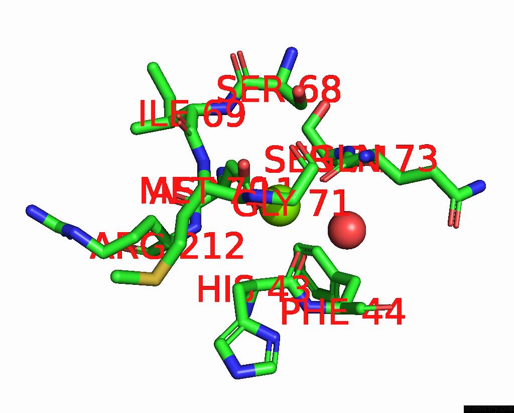
Mono view
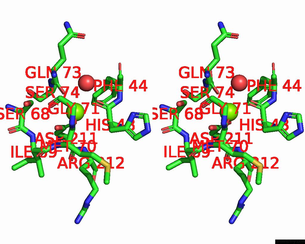
Stereo pair view
Mono view
Stereo pair view
A full contact list of Magnesium with other atoms in the Mg binding
site number 1 of Crystal Structure of Chondroitin Abc Lyase I in Complex with Chondroitin Disaccharide 4,6-Sulfate within 5.0Å range:
|
Reference:
I.Watanabe,
A.Miyanaga,
H.Hoshi,
K.Suzuki,
T.Eguchi.
Biochemical and Crystallographic Assessments of the Effect of 4,6-O-Disulfated Disaccharide Moieties in Chondroitin Sulfate E on Chondroitinase Abc I Activity. Febs J. 2022.
ISSN: ISSN 1742-464X
PubMed: 36478634
DOI: 10.1111/FEBS.16685
Page generated: Thu Oct 3 15:31:14 2024
ISSN: ISSN 1742-464X
PubMed: 36478634
DOI: 10.1111/FEBS.16685
Last articles
Zn in 9J0NZn in 9J0O
Zn in 9J0P
Zn in 9FJX
Zn in 9EKB
Zn in 9C0F
Zn in 9CAH
Zn in 9CH0
Zn in 9CH3
Zn in 9CH1