Magnesium in PDB 7ywm: Crystal Structure of Mycobacterium Abcessus Phosphopantetheine Adenylyltransferase in Complex with Atp
Enzymatic activity of Crystal Structure of Mycobacterium Abcessus Phosphopantetheine Adenylyltransferase in Complex with Atp
All present enzymatic activity of Crystal Structure of Mycobacterium Abcessus Phosphopantetheine Adenylyltransferase in Complex with Atp:
2.7.7.3;
2.7.7.3;
Protein crystallography data
The structure of Crystal Structure of Mycobacterium Abcessus Phosphopantetheine Adenylyltransferase in Complex with Atp, PDB code: 7ywm
was solved by
S.E.Thomas,
A.G.Coyne,
T.L.Blundell,
with X-Ray Crystallography technique. A brief refinement statistics is given in the table below:
| Resolution Low / High (Å) | 64.85 / 1.62 |
| Space group | C 2 2 21 |
| Cell size a, b, c (Å), α, β, γ (°) | 75.827, 125.134, 119.083, 90, 90, 90 |
| R / Rfree (%) | 18.6 / 21.1 |
Magnesium Binding Sites:
The binding sites of Magnesium atom in the Crystal Structure of Mycobacterium Abcessus Phosphopantetheine Adenylyltransferase in Complex with Atp
(pdb code 7ywm). This binding sites where shown within
5.0 Angstroms radius around Magnesium atom.
In total 3 binding sites of Magnesium where determined in the Crystal Structure of Mycobacterium Abcessus Phosphopantetheine Adenylyltransferase in Complex with Atp, PDB code: 7ywm:
Jump to Magnesium binding site number: 1; 2; 3;
In total 3 binding sites of Magnesium where determined in the Crystal Structure of Mycobacterium Abcessus Phosphopantetheine Adenylyltransferase in Complex with Atp, PDB code: 7ywm:
Jump to Magnesium binding site number: 1; 2; 3;
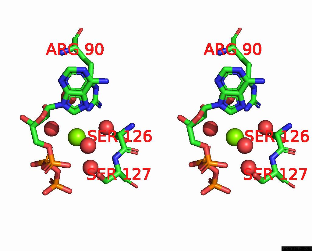
Magnesium binding site 1 out of 3 in 7ywm
Go back to
Magnesium binding site 1 out
of 3 in the Crystal Structure of Mycobacterium Abcessus Phosphopantetheine Adenylyltransferase in Complex with Atp
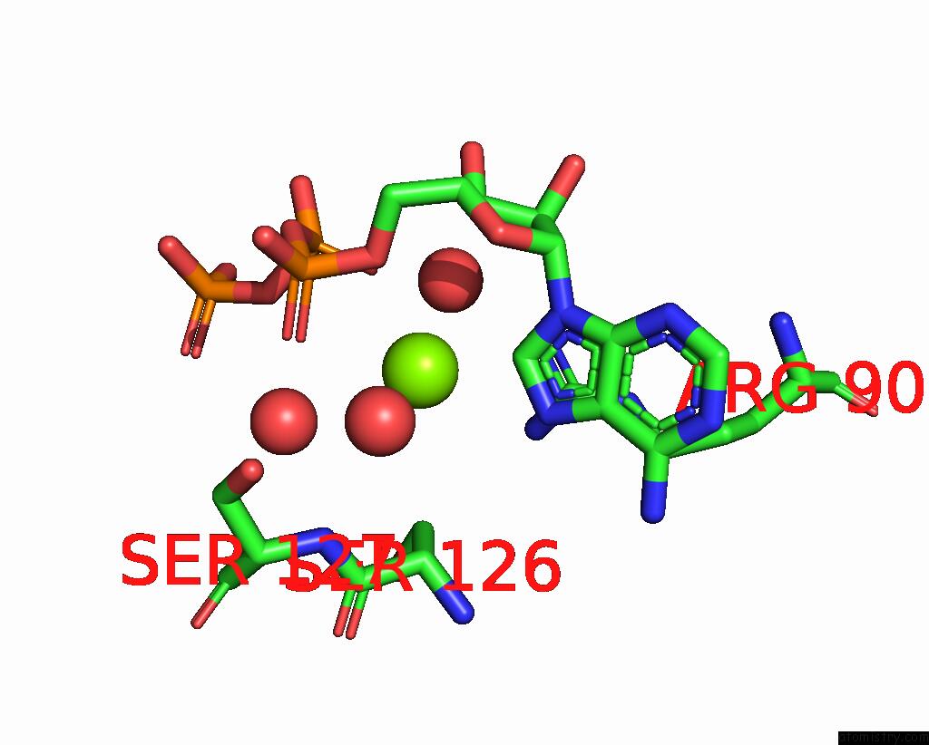
Mono view
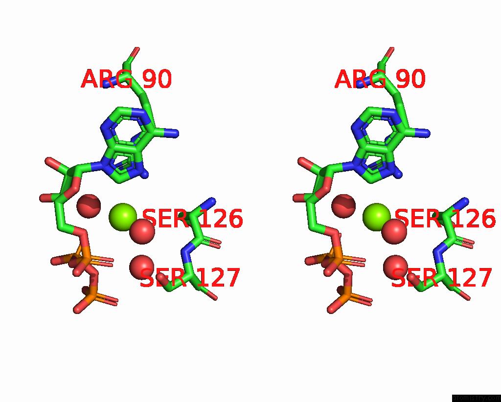
Stereo pair view
Mono view
Stereo pair view
A full contact list of Magnesium with other atoms in the Mg binding
site number 1 of Crystal Structure of Mycobacterium Abcessus Phosphopantetheine Adenylyltransferase in Complex with Atp within 5.0Å range:
|
Magnesium binding site 2 out of 3 in 7ywm
Go back to
Magnesium binding site 2 out
of 3 in the Crystal Structure of Mycobacterium Abcessus Phosphopantetheine Adenylyltransferase in Complex with Atp
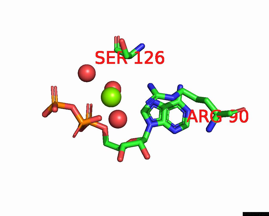
Mono view

Stereo pair view
Mono view
Stereo pair view
A full contact list of Magnesium with other atoms in the Mg binding
site number 2 of Crystal Structure of Mycobacterium Abcessus Phosphopantetheine Adenylyltransferase in Complex with Atp within 5.0Å range:
|
Magnesium binding site 3 out of 3 in 7ywm
Go back to
Magnesium binding site 3 out
of 3 in the Crystal Structure of Mycobacterium Abcessus Phosphopantetheine Adenylyltransferase in Complex with Atp
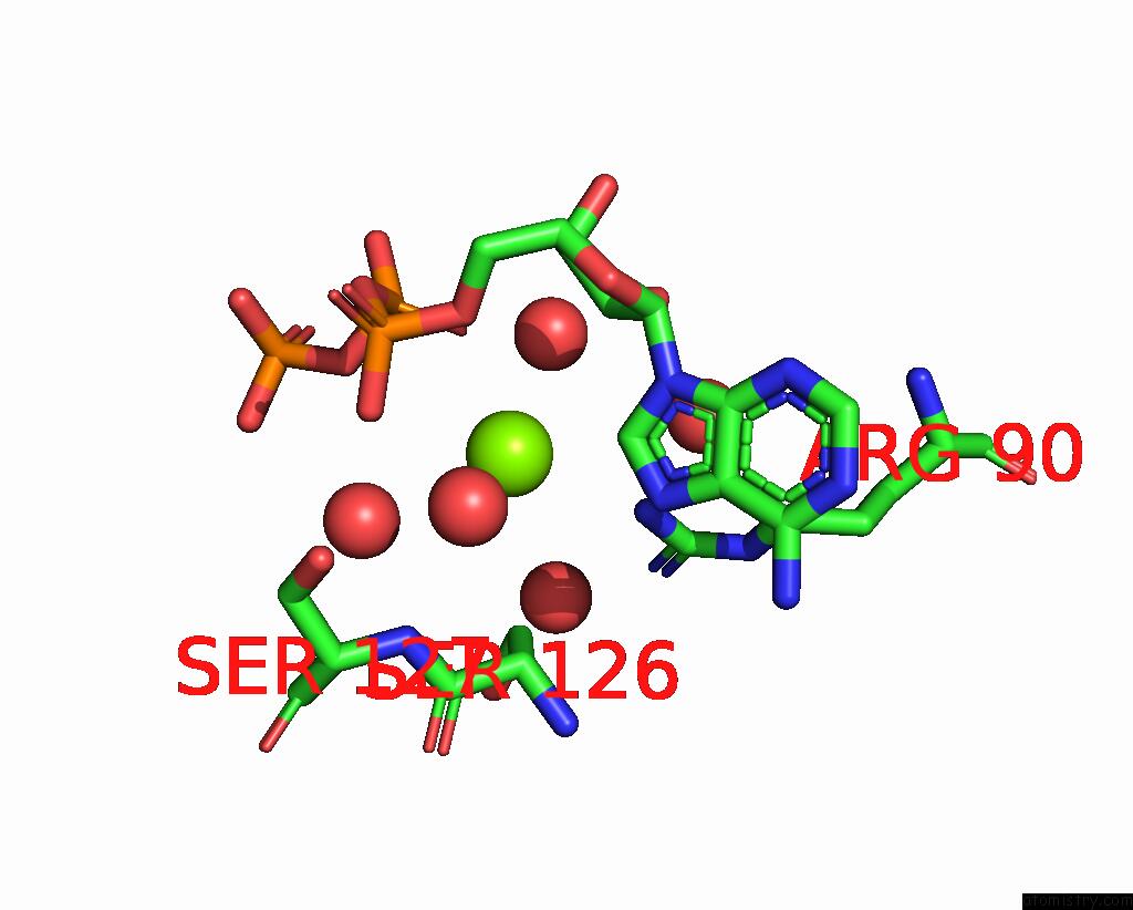
Mono view
Stereo pair view
Mono view
Stereo pair view
A full contact list of Magnesium with other atoms in the Mg binding
site number 3 of Crystal Structure of Mycobacterium Abcessus Phosphopantetheine Adenylyltransferase in Complex with Atp within 5.0Å range:
|
Reference:
S.E.Thomas,
W.J.Mccarthy,
J.El Bakali,
K.P.Brown,
S.Y.Kim,
M.Blaszczyk,
V.Mendes,
C.Abell,
R.A.Floto,
A.G.Coyne,
T.L.Blundell.
Structural Characterization of Mycobacterium Abscessus Phosphopantetheine Adenylyl Transferase Ligand Interactions: Implications For Fragment-Based Drug Design. Front Mol Biosci V. 9 80432 2022.
ISSN: ESSN 2296-889X
PubMed: 35712348
DOI: 10.3389/FMOLB.2022.880432
Page generated: Thu Oct 3 15:43:18 2024
ISSN: ESSN 2296-889X
PubMed: 35712348
DOI: 10.3389/FMOLB.2022.880432
Last articles
Zn in 9JYWZn in 9IR4
Zn in 9IR3
Zn in 9GMX
Zn in 9GMW
Zn in 9JEJ
Zn in 9ERF
Zn in 9ERE
Zn in 9EGV
Zn in 9EGW