Atomistry » Magnesium » PDB 8bdg-8brp » 8beo
Atomistry »
  Magnesium »
    PDB 8bdg-8brp »
      8beo »

Magnesium in PDB 8beo: Crystal Structure of E. Coli Glyoxylate Carboligase Mutant I393A with Map

Enzymatic activity of Crystal Structure of E. Coli Glyoxylate Carboligase Mutant I393A with Map

All present enzymatic activity of Crystal Structure of E. Coli Glyoxylate Carboligase Mutant I393A with Map:
4.1.1.47;

Protein crystallography data

The structure of Crystal Structure of E. Coli Glyoxylate Carboligase Mutant I393A with Map, PDB code: 8beo was solved by B.Shaanan, E.Binshtein, with X-Ray Crystallography technique. A brief refinement statistics is given in the table below:

Resolution Low / High (Å) 48.35 / 1.96
Space group P 41 21 2
Cell size a, b, c (Å), α, β, γ (°) 188.641, 188.641, 246.957, 90, 90, 90
R / Rfree (%) 17.6 / 20.4

Other elements in 8beo:

The structure of Crystal Structure of E. Coli Glyoxylate Carboligase Mutant I393A with Map also contains other interesting chemical elements:

Sodium (Na) 15 atoms
Chlorine (Cl) 1 atom

Magnesium Binding Sites:

Pages:

>>> Page 1 <<< Page 2, Binding sites: 11 - 13;

Binding sites:

The binding sites of Magnesium atom in the Crystal Structure of E. Coli Glyoxylate Carboligase Mutant I393A with Map (pdb code 8beo). This binding sites where shown within 5.0 Angstroms radius around Magnesium atom.
In total 13 binding sites of Magnesium where determined in the Crystal Structure of E. Coli Glyoxylate Carboligase Mutant I393A with Map, PDB code: 8beo:
Jump to Magnesium binding site number: 1; 2; 3; 4; 5; 6; 7; 8; 9; 10;

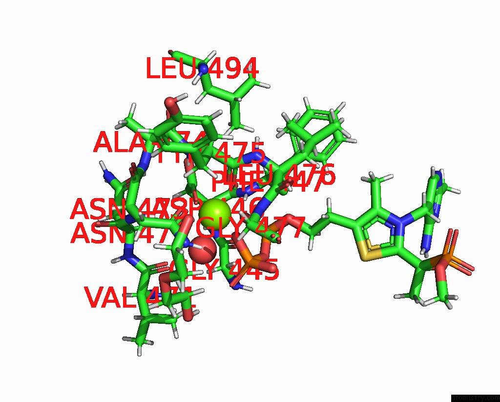
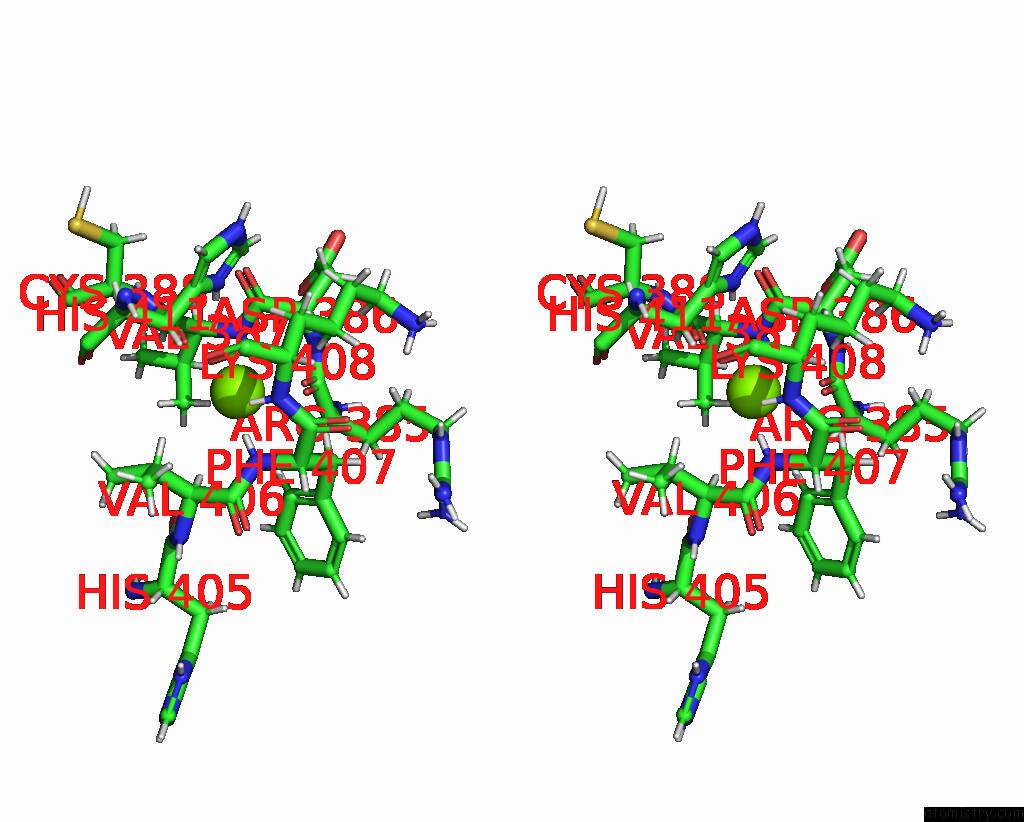
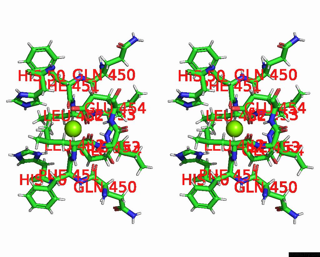
Magnesium binding site 1 out of 13 in 8beo

Go back to Magnesium Binding Sites List in 8beo
Magnesium binding site 1 out of 13 in the Crystal Structure of E. Coli Glyoxylate Carboligase Mutant I393A with Map


Mono view


Stereo pair view

A full contact list of Magnesium with other atoms in the Mg binding site number 1 of Crystal Structure of E. Coli Glyoxylate Carboligase Mutant I393A with Map within 5.0Å range:
probe atom residue distance (Å) B Occ
A:Mg601

b:18.1
occ:1.00
OD1 A:ASN473 2.0 20.7 1.0
O1A A:TDK604 2.0 19.4 1.0
OD1 A:ASP446 2.1 20.1 1.0
O1B A:TDK604 2.1 18.9 1.0
O A:TYR475 2.1 18.3 1.0
O A:HOH880 2.1 17.9 1.0
HD21 A:ASN473 2.9 20.6 1.0
CG A:ASN473 3.0 21.9 1.0
H A:TYR475 3.1 18.3 1.0
H A:ASP446 3.1 20.2 1.0
H A:GLY477 3.2 20.9 1.0
CG A:ASP446 3.2 20.7 1.0
PA A:TDK604 3.3 20.7 1.0
C A:TYR475 3.3 19.1 1.0
PB A:TDK604 3.3 20.1 1.0
ND2 A:ASN473 3.3 20.4 1.0
H A:ASN473 3.4 19.6 1.0
O3A A:TDK604 3.5 19.1 1.0
H A:PHE447 3.7 19.6 1.0
OD2 A:ASP446 3.8 21.6 1.0
N A:TYR475 3.9 18.4 1.0
N A:ASP446 3.9 20.1 1.0
HA3 A:GLY445 3.9 19.5 1.0
O2B A:TDK604 3.9 20.6 1.0
O7 A:TDK604 4.0 21.4 1.0
CA A:TYR475 4.0 18.9 1.0
HA A:LEU476 4.1 19.5 1.0
N A:GLY477 4.1 21.0 1.0
HB2 A:TYR475 4.1 19.1 1.0
HD22 A:ASN473 4.2 21.2 1.0
N A:ASN473 4.2 20.0 1.0
HB3 A:TYR475 4.2 18.9 1.0
CB A:ASN473 4.3 20.5 1.0
N A:LEU476 4.3 19.8 1.0
H11 A:PEG608 4.4 21.8 0.8
CB A:ASP446 4.4 19.9 1.0
CB A:TYR475 4.4 19.1 1.0
HB2 A:PHE447 4.4 19.4 1.0
H A:ALA474 4.5 18.8 1.0
HA2 A:GLY445 4.5 19.7 1.0
HA3 A:GLY477 4.5 21.4 1.0
N A:PHE447 4.5 19.4 1.0
CA A:GLY445 4.5 19.5 1.0
HD11 A:LEU494 4.5 19.6 1.0
O A:VAL471 4.6 19.5 1.0
O2A A:TDK604 4.6 19.5 1.0
HA A:ASN472 4.6 19.2 1.0
CA A:LEU476 4.6 19.3 1.0
O3B A:TDK604 4.6 18.9 1.0
CA A:ASP446 4.6 19.9 1.0
N A:ALA474 4.6 18.8 1.0
CA A:ASN473 4.6 20.5 1.0
C A:GLY445 4.7 19.7 1.0
HB3 A:ASP446 4.7 20.3 1.0
C A:ASN473 4.8 20.3 1.0
H12 A:PEG608 4.8 21.8 0.8
HB3 A:ASN473 4.8 20.9 1.0
C A:LEU476 4.9 20.9 1.0
CA A:GLY477 4.9 21.6 1.0
HB2 A:ASN473 4.9 21.1 1.0
HA A:TYR475 5.0 19.1 1.0

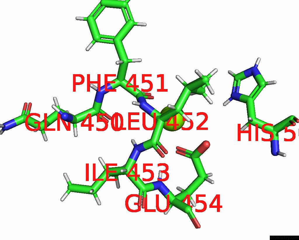
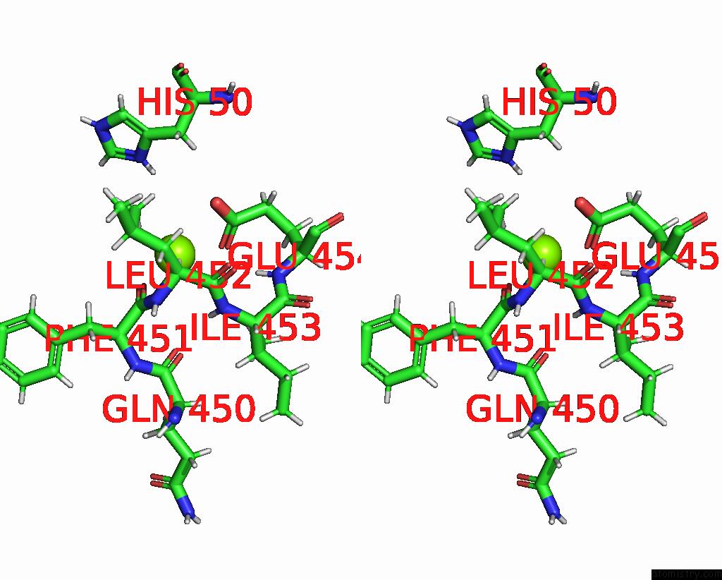
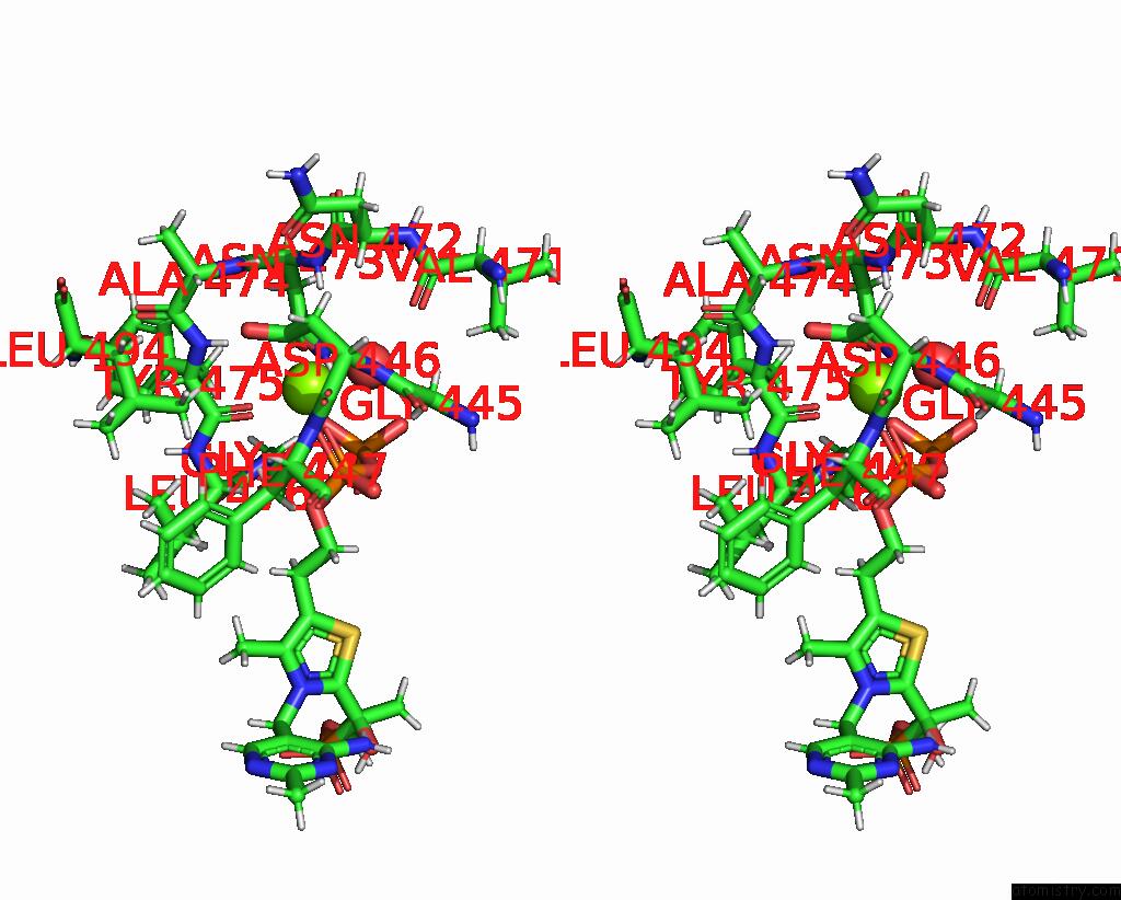
Magnesium binding site 2 out of 13 in 8beo

Go back to Magnesium Binding Sites List in 8beo
Magnesium binding site 2 out of 13 in the Crystal Structure of E. Coli Glyoxylate Carboligase Mutant I393A with Map


Mono view


Stereo pair view

A full contact list of Magnesium with other atoms in the Mg binding site number 2 of Crystal Structure of E. Coli Glyoxylate Carboligase Mutant I393A with Map within 5.0Å range:
probe atom residue distance (Å) B Occ
A:Mg602

b:22.0
occ:0.50
OE1 A:GLU454 2.4 18.7 1.0
O A:PHE451 2.4 17.4 1.0
HA A:LEU452 2.7 17.7 1.0
HD23 A:LEU452 2.9 17.4 1.0
CD A:GLU454 3.3 18.8 1.0
C A:PHE451 3.5 17.5 1.0
OE2 A:GLU454 3.6 18.1 1.0
CA A:LEU452 3.7 17.8 1.0
HD1 A:HIS50 3.7 15.0 0.0
CD2 A:LEU452 3.8 17.3 1.0
H A:ILE453 3.9 18.3 1.0
HD21 A:LEU452 4.0 17.4 1.0
N A:LEU452 4.0 18.1 1.0
H A:GLU454 4.1 18.6 1.0
HG21 A:ILE453 4.4 20.0 1.0
HD22 A:LEU452 4.4 17.3 1.0
C A:LEU452 4.4 17.4 1.0
N A:ILE453 4.4 18.8 1.0
O A:GLN450 4.4 18.1 1.0
ND1 A:HIS50 4.5 17.8 1.0
CG A:GLU454 4.5 18.0 1.0
HG3 A:GLU454 4.6 18.3 1.0
CB A:LEU452 4.7 16.8 1.0
CG A:LEU452 4.7 17.2 1.0
HA A:PHE451 4.7 17.1 1.0
HB3 A:GLU454 4.7 18.3 1.0
CA A:PHE451 4.8 16.6 1.0
HB2 A:HIS50 4.8 17.4 1.0
HG A:LEU452 4.8 17.2 1.0
H A:LEU452 4.9 17.9 1.0
HB3 A:LEU452 4.9 17.1 1.0

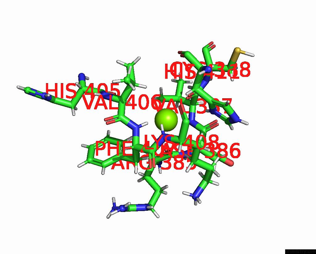
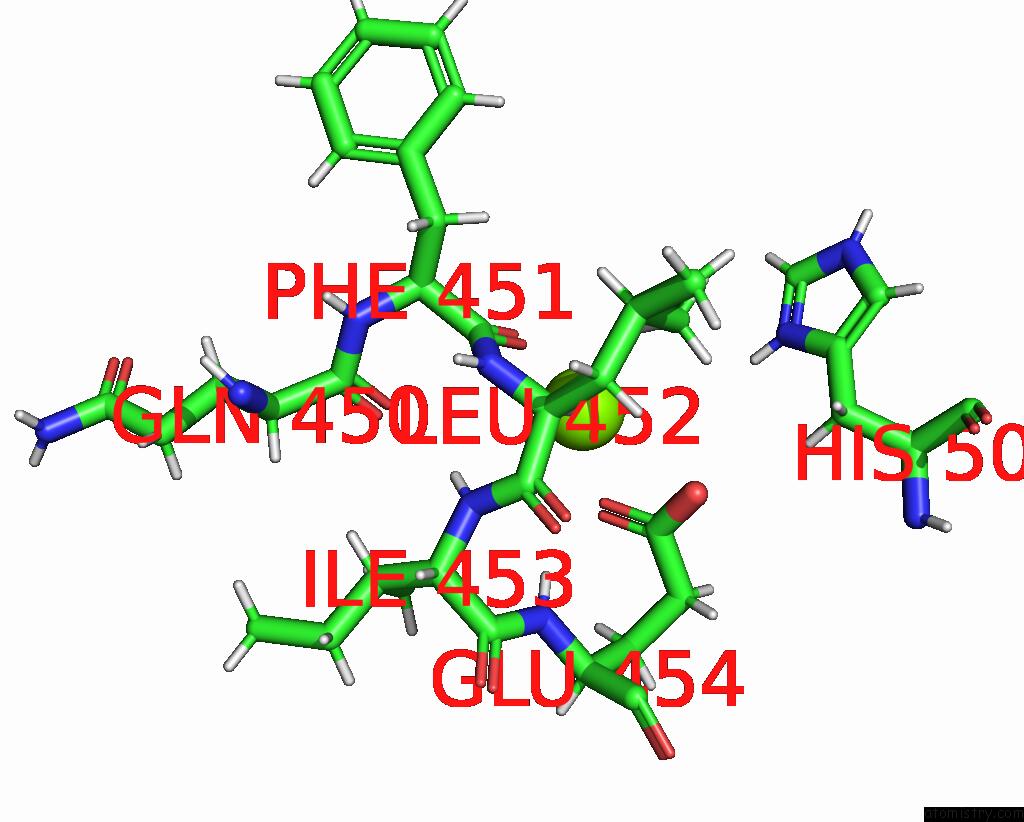
Magnesium binding site 3 out of 13 in 8beo

Go back to Magnesium Binding Sites List in 8beo
Magnesium binding site 3 out of 13 in the Crystal Structure of E. Coli Glyoxylate Carboligase Mutant I393A with Map


Mono view


Stereo pair view

A full contact list of Magnesium with other atoms in the Mg binding site number 3 of Crystal Structure of E. Coli Glyoxylate Carboligase Mutant I393A with Map within 5.0Å range:
probe atom residue distance (Å) B Occ
A:Mg624

b:32.0
occ:1.00
H A:PHE407 2.1 26.3 1.0
O A:ARG385 2.7 27.6 1.0
O A:VAL387 2.9 23.0 1.0
N A:PHE407 2.9 26.7 1.0
HA A:VAL406 3.1 25.9 1.0
HD1 A:PHE407 3.1 23.5 1.0
HB3 A:HIS411 3.1 23.5 1.0
HD1 A:HIS411 3.2 24.5 0.0
HB2 A:PHE407 3.2 25.4 1.0
HB A:VAL406 3.3 28.0 1.0
H A:LYS408 3.3 28.1 1.0
HG3 A:LYS408 3.4 41.5 1.0
HB2 A:HIS411 3.4 23.6 1.0
HG11 A:VAL387 3.5 26.1 1.0
ND1 A:HIS411 3.6 27.4 1.0
CB A:HIS411 3.6 23.5 1.0
C A:ARG385 3.7 26.4 1.0
CA A:VAL406 3.7 26.3 1.0
N A:LYS408 3.8 28.5 1.0
CA A:PHE407 3.8 26.9 1.0
C A:VAL406 3.8 25.9 1.0
HA A:ASP386 3.8 28.0 1.0
CB A:PHE407 3.9 25.4 1.0
CD1 A:PHE407 3.9 24.1 1.0
CG A:HIS411 3.9 24.5 1.0
CB A:VAL406 4.0 27.5 1.0
C A:VAL387 4.0 22.3 1.0
H A:VAL387 4.0 25.7 1.0
N A:VAL387 4.0 25.2 1.0
HB2 A:LYS408 4.1 34.9 1.0
C A:PHE407 4.1 27.5 1.0
C A:ASP386 4.2 26.8 1.0
HG13 A:VAL387 4.2 25.5 1.0
CG1 A:VAL387 4.3 25.9 1.0
CG A:LYS408 4.3 42.0 1.0
CA A:ASP386 4.3 28.4 1.0
CG A:PHE407 4.4 24.4 1.0
HA A:ARG385 4.4 28.4 1.0
N A:ASP386 4.4 26.4 1.0
CB A:LYS408 4.5 35.4 1.0
CA A:VAL387 4.6 24.2 1.0
HG11 A:VAL406 4.6 30.1 1.0
O A:ASP386 4.7 26.5 1.0
CE1 A:HIS411 4.7 27.4 1.0
HA A:PHE407 4.7 26.8 1.0
CA A:ARG385 4.7 28.9 1.0
CA A:LYS408 4.7 29.9 1.0
HB3 A:PHE407 4.7 25.9 1.0
HE2 A:LYS408 4.8 52.4 1.0
HG23 A:VAL406 4.8 27.9 1.0
O A:LYS408 4.8 21.9 1.0
HA A:CYS388 4.8 20.2 1.0
HG2 A:LYS408 4.9 41.9 1.0
CG1 A:VAL406 4.9 31.5 1.0
O A:HIS405 4.9 21.6 1.0
HD2 A:LYS408 4.9 46.6 1.0
HG12 A:VAL387 5.0 26.0 1.0

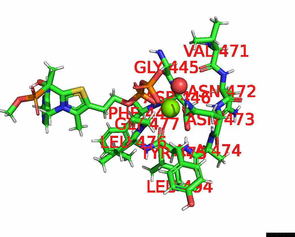
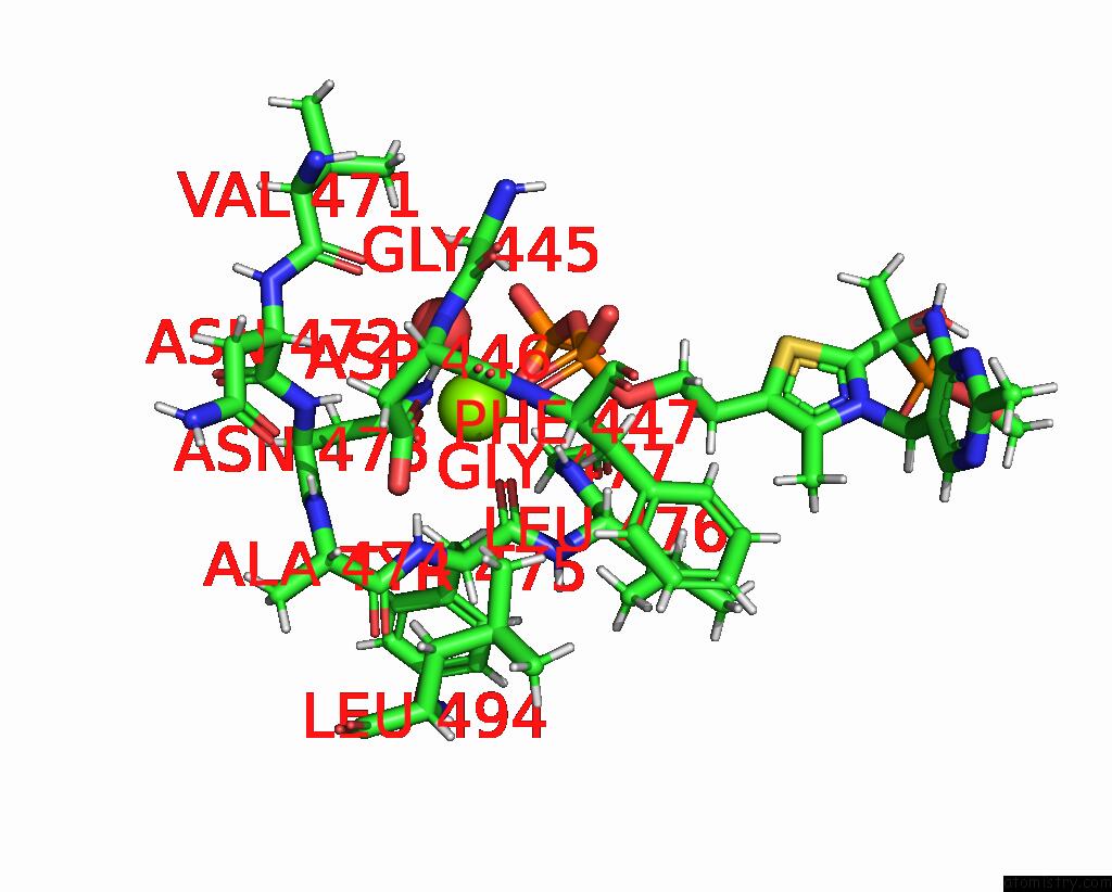
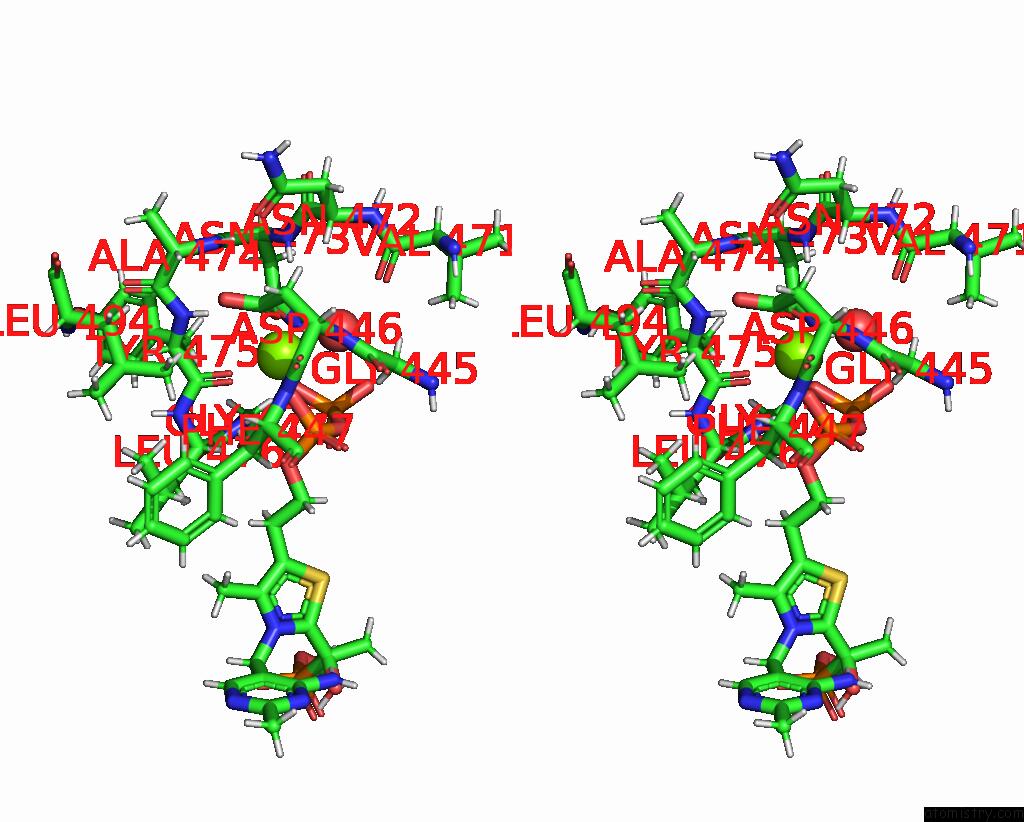
Magnesium binding site 4 out of 13 in 8beo

Go back to Magnesium Binding Sites List in 8beo
Magnesium binding site 4 out of 13 in the Crystal Structure of E. Coli Glyoxylate Carboligase Mutant I393A with Map


Mono view


Stereo pair view

A full contact list of Magnesium with other atoms in the Mg binding site number 4 of Crystal Structure of E. Coli Glyoxylate Carboligase Mutant I393A with Map within 5.0Å range:
probe atom residue distance (Å) B Occ
B:Mg601

b:21.2
occ:1.00
O1A B:TDK604 1.9 22.8 1.0
OD1 B:ASP446 2.1 23.1 1.0
O1B B:TDK604 2.1 21.3 1.0
O B:TYR475 2.1 21.5 1.0
OD1 B:ASN473 2.1 21.8 1.0
O B:HOH837 2.1 21.4 1.0
H B:ASP446 3.0 21.4 1.0
HD21 B:ASN473 3.0 22.1 1.0
CG B:ASN473 3.1 22.5 1.0
H B:TYR475 3.1 22.8 1.0
CG B:ASP446 3.2 22.2 1.0
PA B:TDK604 3.2 23.6 1.0
PB B:TDK604 3.3 22.7 1.0
H B:GLY477 3.3 24.5 1.0
C B:TYR475 3.3 22.3 1.0
H B:ASN473 3.4 22.1 1.0
O3A B:TDK604 3.4 23.8 1.0
ND2 B:ASN473 3.4 22.4 1.0
H B:PHE447 3.5 20.8 1.0
OD2 B:ASP446 3.7 22.4 1.0
HA3 B:GLY445 3.8 21.0 1.0
N B:ASP446 3.8 20.7 1.0
N B:TYR475 3.9 23.1 1.0
O2B B:TDK604 3.9 22.1 1.0
O7 B:TDK604 3.9 25.3 1.0
HA B:LEU476 4.1 22.7 1.0
CA B:TYR475 4.1 23.3 1.0
N B:GLY477 4.1 24.9 1.0
HB2 B:TYR475 4.2 23.2 1.0
HB3 B:TYR475 4.2 22.9 1.0
N B:ASN473 4.2 22.7 1.0
HB2 B:PHE447 4.3 19.9 1.0
HD22 B:ASN473 4.3 22.6 1.0
CB B:ASP446 4.4 21.3 1.0
N B:LEU476 4.4 22.6 1.0
N B:PHE447 4.4 21.0 1.0
CA B:GLY445 4.4 21.0 1.0
HA2 B:GLY445 4.4 21.2 1.0
O2A B:TDK604 4.4 20.1 1.0
CB B:TYR475 4.4 22.9 1.0
CB B:ASN473 4.5 22.1 1.0
H B:ALA474 4.5 23.9 1.0
O B:VAL471 4.5 20.1 1.0
HD11 B:LEU494 4.5 21.3 1.0
O3B B:TDK604 4.5 21.0 1.0
C B:GLY445 4.5 21.7 1.0
CA B:ASP446 4.6 21.3 1.0
HA B:ASN472 4.6 20.7 1.0
HA3 B:GLY477 4.6 25.4 1.0
CA B:LEU476 4.6 22.8 1.0
N B:ALA474 4.7 24.5 1.0
CA B:ASN473 4.7 23.3 1.0
HB3 B:ASP446 4.7 21.8 1.0
C B:ASN473 4.8 24.5 1.0
C B:LEU476 4.9 24.2 1.0
CA B:GLY477 4.9 25.4 1.0
HB2 B:ASN473 5.0 22.9 1.0

Magnesium binding site 5 out of 13 in 8beo

Go back to Magnesium Binding Sites List in 8beo
Magnesium binding site 5 out of 13 in the Crystal Structure of E. Coli Glyoxylate Carboligase Mutant I393A with Map


Mono view


Stereo pair view

A full contact list of Magnesium with other atoms in the Mg binding site number 5 of Crystal Structure of E. Coli Glyoxylate Carboligase Mutant I393A with Map within 5.0Å range:
probe atom residue distance (Å) B Occ
B:Mg602

b:20.8
occ:0.50
O B:PHE451 2.4 19.8 1.0
OE1 B:GLU454 2.4 17.9 1.0
HA B:LEU452 2.8 18.4 1.0
HD23 B:LEU452 3.0 20.9 1.0
CD B:GLU454 3.3 19.0 1.0
C B:PHE451 3.5 18.5 1.0
CA B:LEU452 3.7 18.0 1.0
OE2 B:GLU454 3.7 19.1 1.0
HD1 B:HIS50 3.8 17.9 0.0
CD2 B:LEU452 3.9 21.4 1.0
H B:ILE453 3.9 19.5 1.0
HD21 B:LEU452 3.9 20.9 1.0
N B:LEU452 4.0 17.8 1.0
H B:GLU454 4.0 20.0 1.0
O B:GLN450 4.3 19.8 1.0
HG21 B:ILE453 4.3 19.6 1.0
C B:LEU452 4.4 18.6 1.0
N B:ILE453 4.4 20.1 1.0
HD22 B:LEU452 4.4 20.7 1.0
CG B:GLU454 4.5 18.9 1.0
ND1 B:HIS50 4.6 21.2 1.0
HG3 B:GLU454 4.6 19.3 1.0
HB3 B:GLU454 4.6 20.0 1.0
HA B:PHE451 4.6 18.6 1.0
CB B:LEU452 4.7 18.9 1.0
CA B:PHE451 4.7 18.1 1.0
CG B:LEU452 4.7 19.4 1.0
HB2 B:HIS50 4.8 19.1 1.0
HG B:LEU452 4.9 19.8 1.0
H B:LEU452 4.9 17.9 1.0
N B:GLU454 4.9 20.4 1.0
HB3 B:LEU452 5.0 18.8 1.0

Magnesium binding site 6 out of 13 in 8beo

Go back to Magnesium Binding Sites List in 8beo
Magnesium binding site 6 out of 13 in the Crystal Structure of E. Coli Glyoxylate Carboligase Mutant I393A with Map


Mono view


Stereo pair view

A full contact list of Magnesium with other atoms in the Mg binding site number 6 of Crystal Structure of E. Coli Glyoxylate Carboligase Mutant I393A with Map within 5.0Å range:
probe atom residue distance (Å) B Occ
B:Mg622

b:46.2
occ:1.00
HB3 B:ASN283 2.7 25.2 1.0
HG12 B:VAL311 3.6 30.9 1.0
CB B:ASN283 3.6 25.2 1.0
H B:ASN283 3.9 24.2 1.0
HB2 B:ASN283 4.1 25.4 1.0
CG B:ASN283 4.1 25.2 1.0
HG11 B:VAL311 4.1 31.4 1.0
OD1 B:ASN283 4.2 25.8 1.0
CG1 B:VAL311 4.3 31.7 1.0
O B:HOH843 4.4 29.7 1.0
N B:ASN283 4.4 23.8 1.0
CA B:ASN283 4.5 24.2 1.0
HB B:VAL311 4.6 30.7 1.0
HA B:ASN283 4.6 24.4 1.0

Magnesium binding site 7 out of 13 in 8beo

Go back to Magnesium Binding Sites List in 8beo
Magnesium binding site 7 out of 13 in the Crystal Structure of E. Coli Glyoxylate Carboligase Mutant I393A with Map


Mono view


Stereo pair view

A full contact list of Magnesium with other atoms in the Mg binding site number 7 of Crystal Structure of E. Coli Glyoxylate Carboligase Mutant I393A with Map within 5.0Å range:
probe atom residue distance (Å) B Occ
C:Mg601

b:25.7
occ:1.00
O C:TYR475 2.0 25.1 1.0
OD1 C:ASN473 2.0 28.8 1.0
O1A C:TDK603 2.0 24.9 1.0
O1B C:TDK603 2.1 26.6 1.0
OD1 C:ASP446 2.1 30.0 1.0
O C:HOH862 2.2 25.4 1.0
CG C:ASN473 3.0 29.8 1.0
H C:ASP446 3.1 27.3 1.0
H C:TYR475 3.1 26.3 1.0
HD21 C:ASN473 3.2 30.0 1.0
H C:GLY477 3.2 28.2 1.0
PA C:TDK603 3.2 27.7 1.0
C C:TYR475 3.2 27.0 1.0
CG C:ASP446 3.3 29.3 1.0
PB C:TDK603 3.3 28.8 1.0
ND2 C:ASN473 3.4 30.4 1.0
H C:ASN473 3.4 27.7 1.0
O3A C:TDK603 3.5 27.4 1.0
H C:PHE447 3.6 25.7 1.0
OD2 C:ASP446 3.8 28.6 1.0
N C:TYR475 3.8 26.6 1.0
N C:ASP446 3.9 26.8 1.0
O7 C:TDK603 3.9 29.9 1.0
HA3 C:GLY445 3.9 28.1 1.0
O2B C:TDK603 3.9 27.4 1.0
CA C:TYR475 4.0 27.4 1.0
N C:GLY477 4.0 28.3 1.0
HA C:LEU476 4.0 26.3 1.0
HD22 C:ASN473 4.1 30.4 1.0
HB2 C:TYR475 4.1 28.2 1.0
HB3 C:TYR475 4.1 28.0 1.0
N C:ASN473 4.3 27.5 1.0
N C:LEU476 4.3 26.8 1.0
CB C:TYR475 4.3 28.3 1.0
HB2 C:PHE447 4.4 26.2 1.0
CB C:ASN473 4.4 28.8 1.0
CB C:ASP446 4.4 27.8 1.0
H C:ALA474 4.4 26.7 1.0
HA3 C:GLY477 4.5 30.0 1.0
N C:PHE447 4.5 25.4 1.0
O2A C:TDK603 4.5 26.0 1.0
HD11 C:LEU494 4.5 25.9 1.0
O3B C:TDK603 4.5 25.6 1.0
CA C:LEU476 4.6 26.3 1.0
CA C:GLY445 4.6 28.3 1.0
HA2 C:GLY445 4.6 28.4 1.0
HA C:ASN472 4.6 28.0 1.0
CA C:ASP446 4.6 27.5 1.0
N C:ALA474 4.6 26.6 1.0
O C:VAL471 4.6 25.9 1.0
C C:GLY445 4.7 27.7 1.0
CA C:ASN473 4.7 28.8 1.0
C C:ASN473 4.8 28.4 1.0
HB3 C:ASP446 4.8 28.3 1.0
CA C:GLY477 4.8 30.4 1.0
C C:LEU476 4.8 26.9 1.0
HB3 C:ASN473 4.9 29.2 1.0
HA C:TYR475 4.9 27.5 1.0
HB2 C:ASN473 4.9 29.4 1.0

Magnesium binding site 8 out of 13 in 8beo

Go back to Magnesium Binding Sites List in 8beo
Magnesium binding site 8 out of 13 in the Crystal Structure of E. Coli Glyoxylate Carboligase Mutant I393A with Map


Mono view


Stereo pair view

A full contact list of Magnesium with other atoms in the Mg binding site number 8 of Crystal Structure of E. Coli Glyoxylate Carboligase Mutant I393A with Map within 5.0Å range:
probe atom residue distance (Å) B Occ
C:Mg605

b:22.6
occ:1.00
OE1 E:GLU454 2.3 26.1 1.0
O C:PHE451 2.3 27.0 1.0
OE1 C:GLU454 2.4 27.9 1.0
O E:PHE451 2.4 26.1 1.0
HA C:LEU452 2.7 26.5 1.0
HA E:LEU452 2.8 25.5 1.0
HD23 E:LEU452 2.9 27.2 1.0
HD23 C:LEU452 2.9 26.4 1.0
CD E:GLU454 3.2 27.9 1.0
CD C:GLU454 3.3 29.3 1.0
C C:PHE451 3.4 26.6 1.0
C E:PHE451 3.5 26.5 1.0
CA C:LEU452 3.6 25.9 1.0
OE2 C:GLU454 3.6 28.6 1.0
OE2 E:GLU454 3.7 26.9 1.0
CA E:LEU452 3.7 25.3 1.0
HD1 E:HIS50 3.7 24.4 0.0
HD2 C:HIS50 3.8 24.4 1.0
CD2 E:LEU452 3.8 27.5 1.0
HD21 E:LEU452 3.8 27.1 1.0
CD2 C:LEU452 3.8 26.5 1.0
HD21 C:LEU452 3.9 26.3 1.0
H E:ILE453 3.9 26.3 1.0
H C:ILE453 3.9 27.1 1.0
N C:LEU452 4.0 26.9 1.0
H E:GLU454 4.0 27.2 1.0
N E:LEU452 4.1 25.4 1.0
H C:GLU454 4.1 27.6 1.0
HG21 C:ILE453 4.3 29.0 1.0
HG21 E:ILE453 4.3 27.4 1.0
HD22 E:LEU452 4.3 27.0 1.0
C C:LEU452 4.4 26.4 1.0
C E:LEU452 4.4 25.5 1.0
O C:GLN450 4.4 26.2 1.0
N E:ILE453 4.4 26.5 1.0
N C:ILE453 4.4 27.0 1.0
HD22 C:LEU452 4.4 26.2 1.0
CG E:GLU454 4.4 27.7 1.0
O E:GLN450 4.5 25.1 1.0
ND1 E:HIS50 4.5 29.6 1.0
CG C:GLU454 4.5 28.6 1.0
HB3 E:GLU454 4.5 27.6 1.0
HG3 E:GLU454 4.5 27.7 1.0
HG3 C:GLU454 4.5 28.9 1.0
CD2 C:HIS50 4.6 24.4 1.0
HA C:PHE451 4.6 26.9 1.0
CB C:LEU452 4.7 26.7 1.0
HB3 C:GLU454 4.7 28.3 1.0
CB E:LEU452 4.7 25.7 1.0
HA E:PHE451 4.7 26.4 1.0
CG C:LEU452 4.7 25.6 1.0
CA C:PHE451 4.7 26.9 1.0
CG E:LEU452 4.7 26.2 1.0
CA E:PHE451 4.8 25.8 1.0
HB2 E:HIS50 4.8 26.1 1.0
HG C:LEU452 4.8 25.9 1.0
HG E:LEU452 4.8 26.4 1.0
HB2 C:HIS50 4.9 26.1 1.0
H C:LEU452 4.9 26.5 1.0
N E:GLU454 4.9 27.4 1.0
HB3 C:LEU452 4.9 26.3 1.0
HB3 E:LEU452 4.9 25.7 1.0
H E:LEU452 5.0 25.6 1.0
N C:GLU454 5.0 27.9 1.0

Magnesium binding site 9 out of 13 in 8beo

Go back to Magnesium Binding Sites List in 8beo
Magnesium binding site 9 out of 13 in the Crystal Structure of E. Coli Glyoxylate Carboligase Mutant I393A with Map


Mono view


Stereo pair view

A full contact list of Magnesium with other atoms in the Mg binding site number 9 of Crystal Structure of E. Coli Glyoxylate Carboligase Mutant I393A with Map within 5.0Å range:
probe atom residue distance (Å) B Occ
D:Mg601

b:28.0
occ:1.00
O1B D:TDK603 2.0 27.2 1.0
O D:TYR475 2.1 27.0 1.0
O1A D:TDK603 2.1 28.5 1.0
OD1 D:ASP446 2.1 28.1 1.0
OD1 D:ASN473 2.2 30.6 1.0
O D:HOH834 2.2 26.2 1.0
CG D:ASN473 3.0 31.6 1.0
HD21 D:ASN473 3.1 31.4 1.0
H D:ASP446 3.1 29.1 1.0
H D:TYR475 3.2 28.4 1.0
H D:GLY477 3.2 27.6 1.0
PB D:TDK603 3.2 27.6 1.0
CG D:ASP446 3.3 30.2 1.0
PA D:TDK603 3.3 29.2 1.0
C D:TYR475 3.3 28.8 1.0
ND2 D:ASN473 3.3 31.8 1.0
H D:ASN473 3.4 30.3 1.0
O3A D:TDK603 3.5 28.7 1.0
H D:PHE447 3.6 29.6 1.0
OD2 D:ASP446 3.8 30.1 1.0
HA3 D:GLY445 3.8 30.0 1.0
N D:ASP446 3.9 28.1 1.0
N D:TYR475 3.9 28.4 1.0
O2B D:TDK603 3.9 29.1 1.0
O7 D:TDK603 4.0 31.4 1.0
N D:GLY477 4.1 27.4 1.0
HA D:LEU476 4.1 28.5 1.0
CA D:TYR475 4.1 30.0 1.0
HD22 D:ASN473 4.1 31.9 1.0
HB2 D:TYR475 4.2 30.9 1.0
HB3 D:TYR475 4.2 30.6 1.0
N D:ASN473 4.3 30.9 1.0
N D:LEU476 4.3 28.9 1.0
CB D:ASN473 4.4 30.8 1.0
O3B D:TDK603 4.4 27.4 1.0
HB2 D:PHE447 4.4 29.6 1.0
CB D:TYR475 4.4 30.7 1.0
CB D:ASP446 4.4 30.2 1.0
HA2 D:GLY445 4.5 30.2 1.0
CA D:GLY445 4.5 30.1 1.0
HA3 D:GLY477 4.5 28.5 1.0
N D:PHE447 4.5 29.3 1.0
H D:ALA474 4.5 29.5 1.0
HA D:ASN472 4.6 29.6 1.0
O2A D:TDK603 4.6 28.1 1.0
HD11 D:LEU494 4.6 31.0 1.0
O D:VAL471 4.6 29.5 1.0
C D:GLY445 4.6 29.8 1.0
CA D:LEU476 4.6 28.8 1.0
CA D:ASP446 4.6 29.2 1.0
N D:ALA474 4.7 29.6 1.0
CA D:ASN473 4.7 30.4 1.0
HB3 D:ASP446 4.8 30.2 1.0
C D:ASN473 4.8 30.8 1.0
CA D:GLY477 4.9 28.5 1.0
C D:LEU476 4.9 28.6 1.0
HB3 D:ASN473 4.9 31.0 1.0
HB2 D:ASN473 5.0 31.4 1.0
HA D:TYR475 5.0 29.9 1.0

Magnesium binding site 10 out of 13 in 8beo

Go back to Magnesium Binding Sites List in 8beo
Magnesium binding site 10 out of 13 in the Crystal Structure of E. Coli Glyoxylate Carboligase Mutant I393A with Map


Mono view


Stereo pair view

A full contact list of Magnesium with other atoms in the Mg binding site number 10 of Crystal Structure of E. Coli Glyoxylate Carboligase Mutant I393A with Map within 5.0Å range:
probe atom residue distance (Å) B Occ
E:Mg601

b:29.5
occ:1.00
O E:TYR475 2.0 26.0 1.0
O1B E:TDK603 2.1 31.0 1.0
O1A E:TDK603 2.1 32.9 1.0
OD1 E:ASP446 2.1 31.2 1.0
OD1 E:ASN473 2.1 28.7 1.0
O E:HOH733 2.3 24.7 1.0
CG E:ASN473 3.1 30.9 1.0
H E:TYR475 3.1 29.9 1.0
HD21 E:ASN473 3.1 31.6 1.0
H E:ASP446 3.1 30.9 1.0
PA E:TDK603 3.2 32.3 1.0
CG E:ASP446 3.2 30.8 1.0
C E:TYR475 3.2 28.6 1.0
PB E:TDK603 3.2 33.0 1.0
H E:GLY477 3.3 32.0 1.0
ND2 E:ASN473 3.4 32.2 1.0
O3A E:TDK603 3.4 34.0 1.0
H E:ASN473 3.5 29.5 1.0
H E:PHE447 3.6 28.1 1.0
OD2 E:ASP446 3.8 30.2 1.0
N E:TYR475 3.8 30.3 1.0
N E:ASP446 3.8 31.1 1.0
HA3 E:GLY445 3.9 30.3 1.0
O7 E:TDK603 3.9 36.7 1.0
O2B E:TDK603 3.9 30.4 1.0
CA E:TYR475 4.0 29.5 1.0
HA E:LEU476 4.1 29.5 1.0
N E:GLY477 4.1 32.4 1.0
HB2 E:TYR475 4.1 29.4 1.0
HD22 E:ASN473 4.2 32.1 1.0
HB3 E:TYR475 4.2 29.2 1.0
HB2 E:PHE447 4.3 26.8 1.0
N E:LEU476 4.3 28.8 1.0
N E:ASN473 4.3 29.6 1.0
CB E:TYR475 4.4 29.1 1.0
CB E:ASP446 4.4 30.1 1.0
N E:PHE447 4.4 27.8 1.0
CB E:ASN473 4.5 30.7 1.0
O2A E:TDK603 4.5 31.7 1.0
HA2 E:GLY445 4.5 30.5 1.0
CA E:GLY445 4.5 30.8 1.0
HD11 E:LEU494 4.5 32.2 1.0
HA3 E:GLY477 4.5 33.4 1.0
O3B E:TDK603 4.5 30.7 1.0
H E:ALA474 4.5 29.2 1.0
O E:VAL471 4.6 28.4 1.0
C E:GLY445 4.6 30.5 1.0
CA E:ASP446 4.6 29.7 1.0
CA E:LEU476 4.6 29.9 1.0
HA E:ASN472 4.6 29.4 1.0
N E:ALA474 4.7 28.9 1.0
CA E:ASN473 4.8 30.2 1.0
HB3 E:ASP446 4.8 30.4 1.0
C E:ASN473 4.8 29.8 1.0
C E:LEU476 4.9 31.3 1.0
CA E:GLY477 4.9 33.5 1.0
HA E:TYR475 4.9 29.6 1.0
HB3 E:ASN473 5.0 30.8 1.0

Reference:

B.Shaanan, E.Binshtein. Crystal Structure of E. Coli Glyoxylate Carboligase Mutant I393A with Map To Be Published.
Page generated: Thu Dec 28 08:12:55 2023

Last articles

Zn in 8WB0
Zn in 8WAX
Zn in 8WAU
Zn in 8WAZ
Zn in 8WAY
Zn in 8WAV
Zn in 8WAW
Zn in 8WAT
Zn in 8W7M
Zn in 8WD3
© Copyright 2008-2020 by atomistry.com
Home   |    Site Map   |    Copyright   |    Contact us   |    Privacy