Magnesium in PDB 8i4q: Crystal Structure of 6-Phosphogluconate Dehydrogenase From Corynebacterium Glutamicum
Protein crystallography data
The structure of Crystal Structure of 6-Phosphogluconate Dehydrogenase From Corynebacterium Glutamicum, PDB code: 8i4q
was solved by
H.Yu,
K.-J.Kim,
with X-Ray Crystallography technique. A brief refinement statistics is given in the table below:
| Resolution Low / High (Å) | 33.84 / 1.90 |
| Space group | P 21 21 21 |
| Cell size a, b, c (Å), α, β, γ (°) | 64.004, 119.471, 153.446, 90, 90, 90 |
| R / Rfree (%) | 16.4 / 20.2 |
Magnesium Binding Sites:
The binding sites of Magnesium atom in the Crystal Structure of 6-Phosphogluconate Dehydrogenase From Corynebacterium Glutamicum
(pdb code 8i4q). This binding sites where shown within
5.0 Angstroms radius around Magnesium atom.
In total 7 binding sites of Magnesium where determined in the Crystal Structure of 6-Phosphogluconate Dehydrogenase From Corynebacterium Glutamicum, PDB code: 8i4q:
Jump to Magnesium binding site number: 1; 2; 3; 4; 5; 6; 7;
In total 7 binding sites of Magnesium where determined in the Crystal Structure of 6-Phosphogluconate Dehydrogenase From Corynebacterium Glutamicum, PDB code: 8i4q:
Jump to Magnesium binding site number: 1; 2; 3; 4; 5; 6; 7;
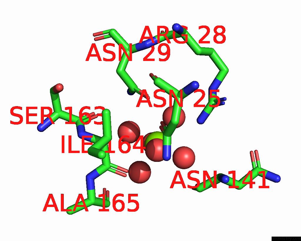
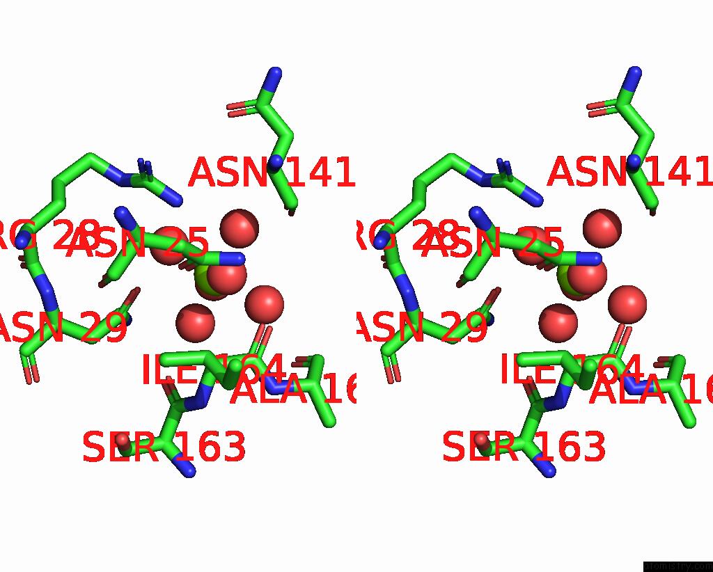
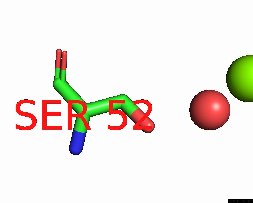
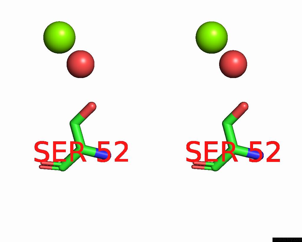
Magnesium binding site 1 out of 7 in 8i4q
Go back to
Magnesium binding site 1 out
of 7 in the Crystal Structure of 6-Phosphogluconate Dehydrogenase From Corynebacterium Glutamicum

Mono view
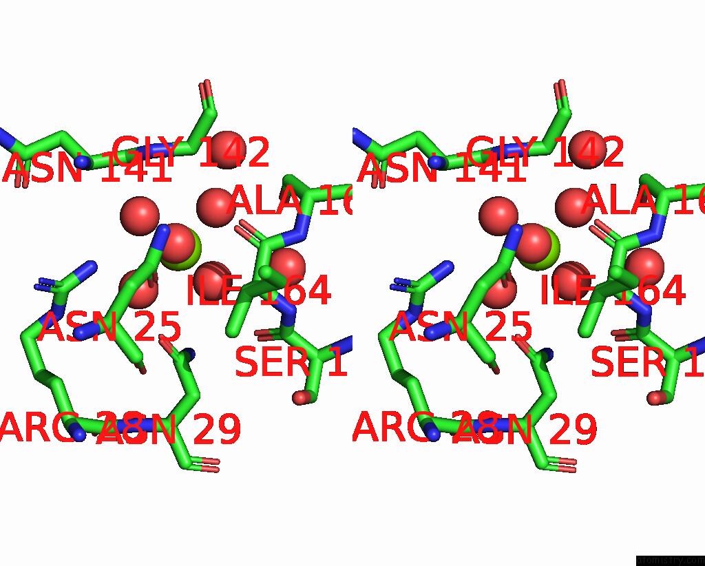
Stereo pair view
Mono view
Stereo pair view
A full contact list of Magnesium with other atoms in the Mg binding
site number 1 of Crystal Structure of 6-Phosphogluconate Dehydrogenase From Corynebacterium Glutamicum within 5.0Å range:
|
Magnesium binding site 2 out of 7 in 8i4q
Go back to
Magnesium binding site 2 out
of 7 in the Crystal Structure of 6-Phosphogluconate Dehydrogenase From Corynebacterium Glutamicum
Mono view

Stereo pair view
Mono view
Stereo pair view
A full contact list of Magnesium with other atoms in the Mg binding
site number 2 of Crystal Structure of 6-Phosphogluconate Dehydrogenase From Corynebacterium Glutamicum within 5.0Å range:
|
Magnesium binding site 3 out of 7 in 8i4q
Go back to
Magnesium binding site 3 out
of 7 in the Crystal Structure of 6-Phosphogluconate Dehydrogenase From Corynebacterium Glutamicum
Mono view
Stereo pair view
Mono view
Stereo pair view
A full contact list of Magnesium with other atoms in the Mg binding
site number 3 of Crystal Structure of 6-Phosphogluconate Dehydrogenase From Corynebacterium Glutamicum within 5.0Å range:
|
Magnesium binding site 4 out of 7 in 8i4q
Go back to
Magnesium binding site 4 out
of 7 in the Crystal Structure of 6-Phosphogluconate Dehydrogenase From Corynebacterium Glutamicum
Mono view
Stereo pair view
Mono view
Stereo pair view
A full contact list of Magnesium with other atoms in the Mg binding
site number 4 of Crystal Structure of 6-Phosphogluconate Dehydrogenase From Corynebacterium Glutamicum within 5.0Å range:
|
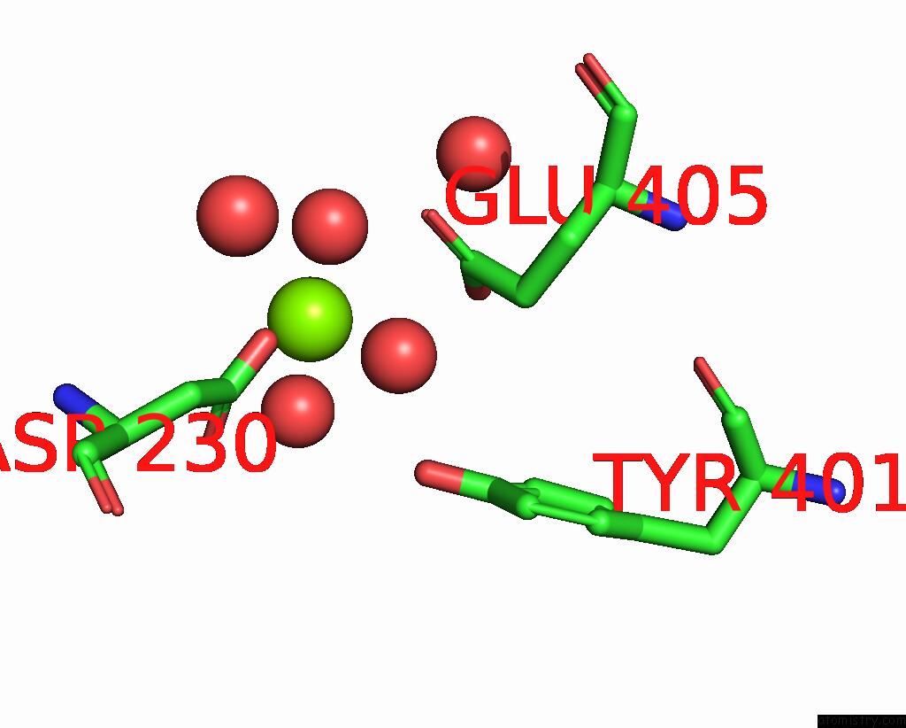
Magnesium binding site 5 out of 7 in 8i4q
Go back to
Magnesium binding site 5 out
of 7 in the Crystal Structure of 6-Phosphogluconate Dehydrogenase From Corynebacterium Glutamicum
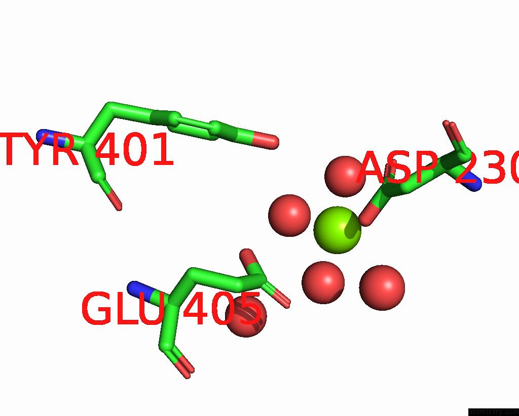
Mono view
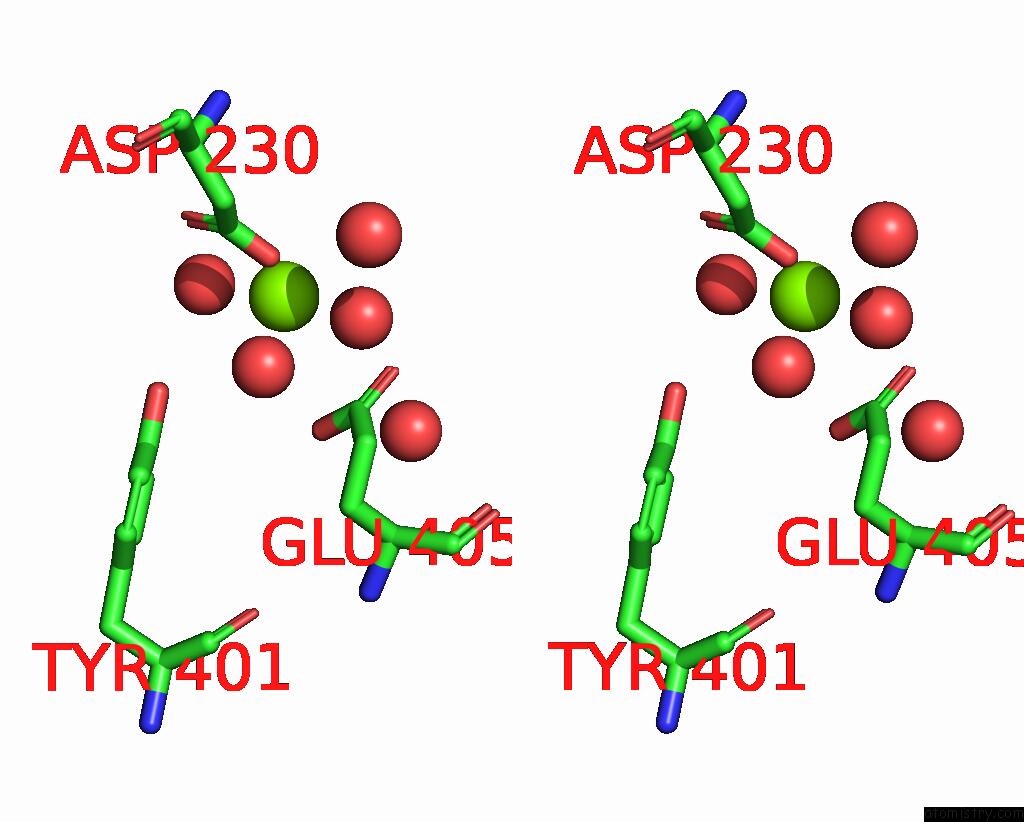
Stereo pair view
Mono view
Stereo pair view
A full contact list of Magnesium with other atoms in the Mg binding
site number 5 of Crystal Structure of 6-Phosphogluconate Dehydrogenase From Corynebacterium Glutamicum within 5.0Å range:
|
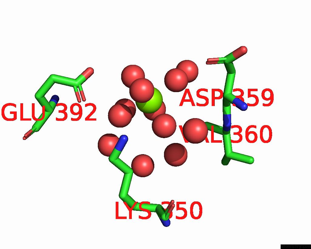
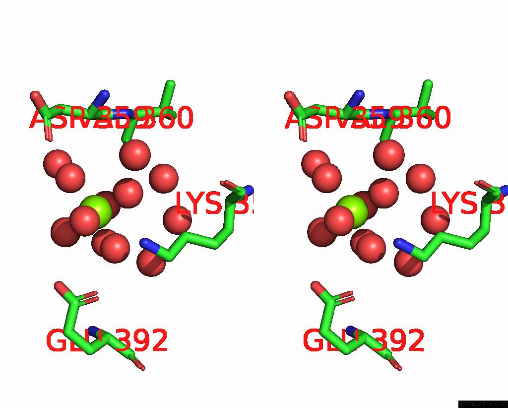
Magnesium binding site 6 out of 7 in 8i4q
Go back to
Magnesium binding site 6 out
of 7 in the Crystal Structure of 6-Phosphogluconate Dehydrogenase From Corynebacterium Glutamicum
Mono view
Stereo pair view
Mono view
Stereo pair view
A full contact list of Magnesium with other atoms in the Mg binding
site number 6 of Crystal Structure of 6-Phosphogluconate Dehydrogenase From Corynebacterium Glutamicum within 5.0Å range:
|
Magnesium binding site 7 out of 7 in 8i4q
Go back to
Magnesium binding site 7 out
of 7 in the Crystal Structure of 6-Phosphogluconate Dehydrogenase From Corynebacterium Glutamicum
Mono view
Stereo pair view
Mono view
Stereo pair view
| A full contact list of Magnesium with other atoms in the Mg binding site number 7 of Crystal Structure of 6-Phosphogluconate Dehydrogenase From Corynebacterium Glutamicum within 5.0Å range: |
Reference:
H.Yu,
J.Hong,
J.Seok,
Y.B.Seu,
I.K.Kim,
K.J.Kim.
Crystal Structures of 6-Phosphogluconate Dehydrogenase From Corynebacterium Glutamicum. J Microbiol Biotechnol. V. 33 1 2023.
ISSN: ESSN 1738-8872
PubMed: 37417004
DOI: 10.4014/JMB.2305.05002
Page generated: Fri Oct 4 09:06:24 2024
ISSN: ESSN 1738-8872
PubMed: 37417004
DOI: 10.4014/JMB.2305.05002
Last articles
K in 7G79K in 7G76
K in 7G7D
K in 7G71
K in 7G74
K in 7G7F
K in 7G77
K in 7G73
K in 7G78
K in 7G7H