Magnesium in PDB 8j3o: Formate Dehydrogenase Wild-Type Enzyme From Candida Dubliniensis Complexed with Nadh
Enzymatic activity of Formate Dehydrogenase Wild-Type Enzyme From Candida Dubliniensis Complexed with Nadh
All present enzymatic activity of Formate Dehydrogenase Wild-Type Enzyme From Candida Dubliniensis Complexed with Nadh:
1.17.1.9;
1.17.1.9;
Protein crystallography data
The structure of Formate Dehydrogenase Wild-Type Enzyme From Candida Dubliniensis Complexed with Nadh, PDB code: 8j3o
was solved by
W.Ma,
Y.C.Zheng,
Q.Geng,
C.Chen,
with X-Ray Crystallography technique. A brief refinement statistics is given in the table below:
| Resolution Low / High (Å) | 44.58 / 2.65 |
| Space group | P 1 |
| Cell size a, b, c (Å), α, β, γ (°) | 51.402, 60.156, 120.482, 90, 90, 111.02 |
| R / Rfree (%) | 22.6 / 27.8 |
Magnesium Binding Sites:
The binding sites of Magnesium atom in the Formate Dehydrogenase Wild-Type Enzyme From Candida Dubliniensis Complexed with Nadh
(pdb code 8j3o). This binding sites where shown within
5.0 Angstroms radius around Magnesium atom.
In total 3 binding sites of Magnesium where determined in the Formate Dehydrogenase Wild-Type Enzyme From Candida Dubliniensis Complexed with Nadh, PDB code: 8j3o:
Jump to Magnesium binding site number: 1; 2; 3;
In total 3 binding sites of Magnesium where determined in the Formate Dehydrogenase Wild-Type Enzyme From Candida Dubliniensis Complexed with Nadh, PDB code: 8j3o:
Jump to Magnesium binding site number: 1; 2; 3;
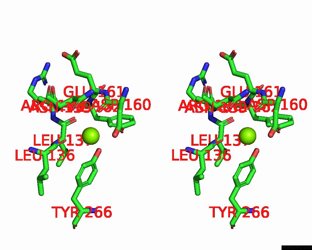
Magnesium binding site 1 out of 3 in 8j3o
Go back to
Magnesium binding site 1 out
of 3 in the Formate Dehydrogenase Wild-Type Enzyme From Candida Dubliniensis Complexed with Nadh

Mono view
Stereo pair view
Mono view
Stereo pair view
A full contact list of Magnesium with other atoms in the Mg binding
site number 1 of Formate Dehydrogenase Wild-Type Enzyme From Candida Dubliniensis Complexed with Nadh within 5.0Å range:
|
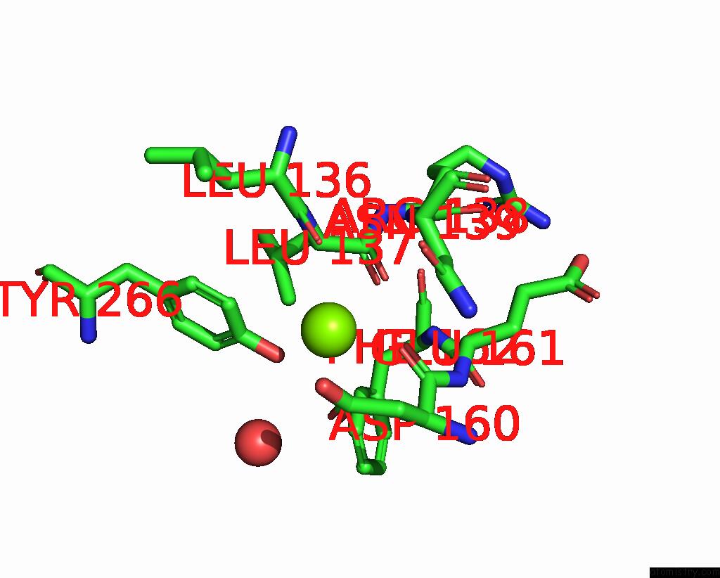
Magnesium binding site 2 out of 3 in 8j3o
Go back to
Magnesium binding site 2 out
of 3 in the Formate Dehydrogenase Wild-Type Enzyme From Candida Dubliniensis Complexed with Nadh
Mono view

Stereo pair view
Mono view
Stereo pair view
A full contact list of Magnesium with other atoms in the Mg binding
site number 2 of Formate Dehydrogenase Wild-Type Enzyme From Candida Dubliniensis Complexed with Nadh within 5.0Å range:
|
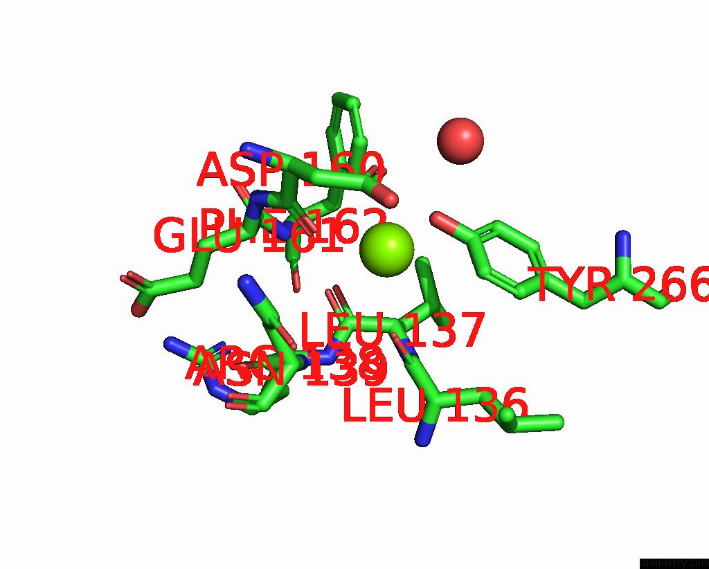
Magnesium binding site 3 out of 3 in 8j3o
Go back to
Magnesium binding site 3 out
of 3 in the Formate Dehydrogenase Wild-Type Enzyme From Candida Dubliniensis Complexed with Nadh
Mono view

Stereo pair view
Mono view
Stereo pair view
A full contact list of Magnesium with other atoms in the Mg binding
site number 3 of Formate Dehydrogenase Wild-Type Enzyme From Candida Dubliniensis Complexed with Nadh within 5.0Å range:
|
Reference:
W.Ma,
Q.Geng,
C.Chen,
Y.C.Zheng,
H.L.Yu,
J.H.Xu.
Engineering A Formate Dehydrogenase For Nadph Regeneration. Chembiochem 00390 2023.
ISSN: ESSN 1439-7633
PubMed: 37455264
DOI: 10.1002/CBIC.202300390
Page generated: Fri Oct 4 11:30:05 2024
ISSN: ESSN 1439-7633
PubMed: 37455264
DOI: 10.1002/CBIC.202300390
Last articles
Zn in 9JYWZn in 9IR4
Zn in 9IR3
Zn in 9GMX
Zn in 9GMW
Zn in 9JEJ
Zn in 9ERF
Zn in 9ERE
Zn in 9EGV
Zn in 9EGW