Magnesium in PDB 8pdd: Thioredoxin Glutathione Reductase of Schistosoma Mansoni at 1.25A Resolution.
Enzymatic activity of Thioredoxin Glutathione Reductase of Schistosoma Mansoni at 1.25A Resolution.
All present enzymatic activity of Thioredoxin Glutathione Reductase of Schistosoma Mansoni at 1.25A Resolution.:
1.6.4.5;
1.6.4.5;
Protein crystallography data
The structure of Thioredoxin Glutathione Reductase of Schistosoma Mansoni at 1.25A Resolution., PDB code: 8pdd
was solved by
L.Ribeiro,
B.O.Montoya,
J.T.Moreira-Filho,
S.Bowyer,
A.Verma,
B.J.Neves,
R.J.Owens,
C.H.Andrade,
F.P.Silva-Jr,
N.Furnham,
with X-Ray Crystallography technique. A brief refinement statistics is given in the table below:
| Resolution Low / High (Å) | 61.85 / 1.25 |
| Space group | P 1 21 1 |
| Cell size a, b, c (Å), α, β, γ (°) | 61.9, 102.4, 131.46, 90, 92.39, 90 |
| R / Rfree (%) | 14.3 / 16.3 |
Magnesium Binding Sites:
The binding sites of Magnesium atom in the Thioredoxin Glutathione Reductase of Schistosoma Mansoni at 1.25A Resolution.
(pdb code 8pdd). This binding sites where shown within
5.0 Angstroms radius around Magnesium atom.
In total 3 binding sites of Magnesium where determined in the Thioredoxin Glutathione Reductase of Schistosoma Mansoni at 1.25A Resolution., PDB code: 8pdd:
Jump to Magnesium binding site number: 1; 2; 3;
In total 3 binding sites of Magnesium where determined in the Thioredoxin Glutathione Reductase of Schistosoma Mansoni at 1.25A Resolution., PDB code: 8pdd:
Jump to Magnesium binding site number: 1; 2; 3;
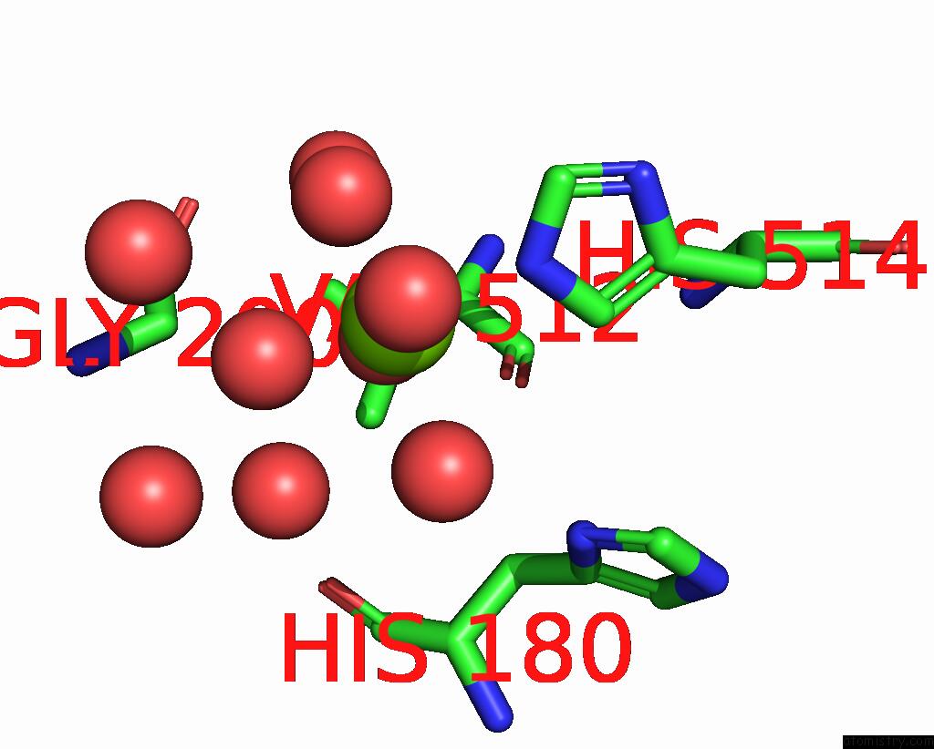
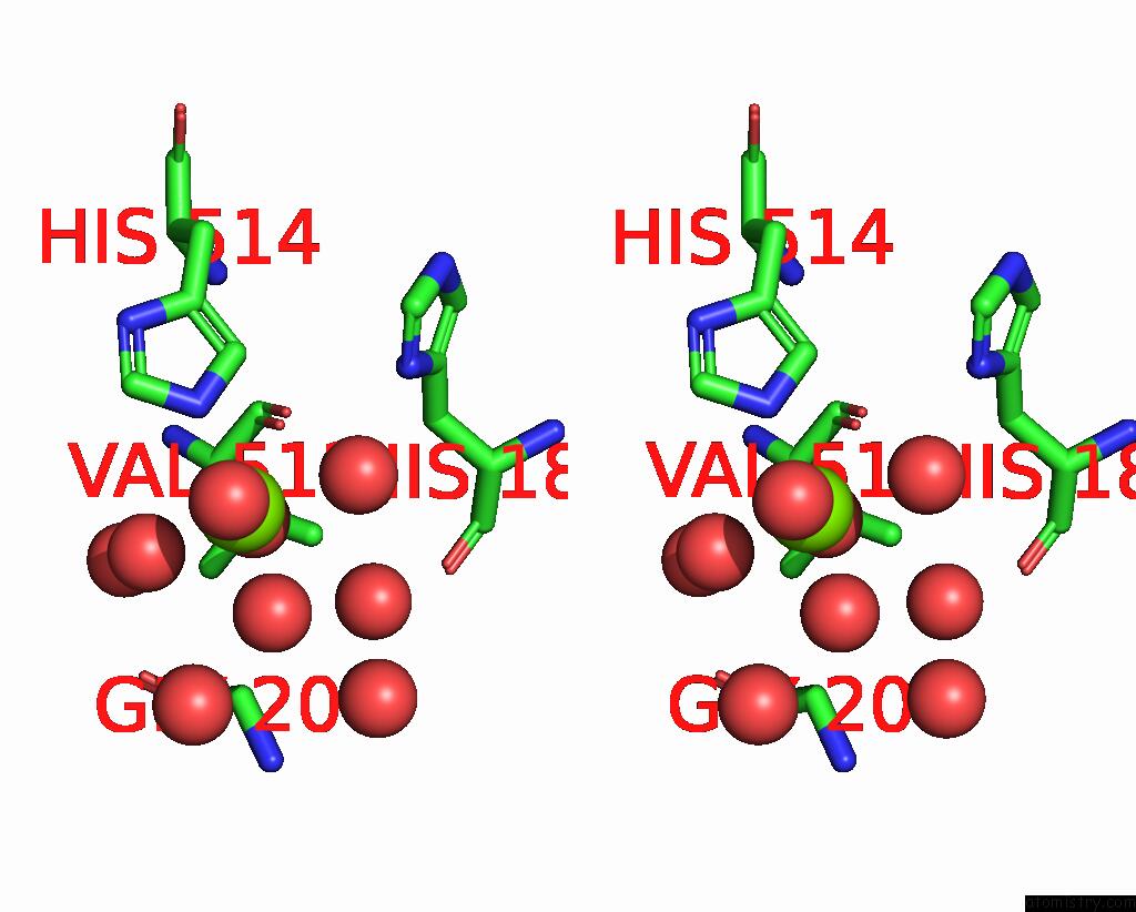
Magnesium binding site 1 out of 3 in 8pdd
Go back to
Magnesium binding site 1 out
of 3 in the Thioredoxin Glutathione Reductase of Schistosoma Mansoni at 1.25A Resolution.
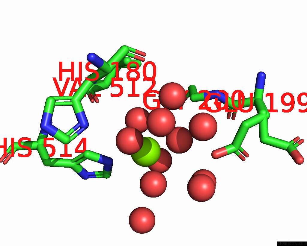
Mono view
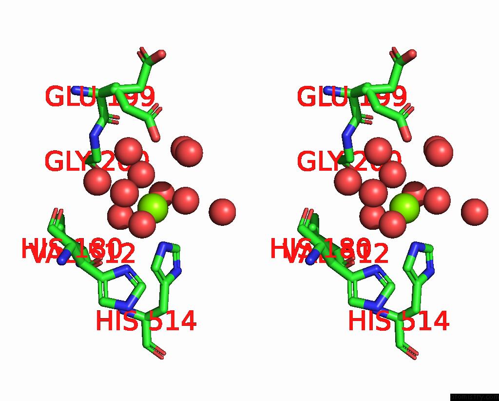
Stereo pair view
Mono view
Stereo pair view
A full contact list of Magnesium with other atoms in the Mg binding
site number 1 of Thioredoxin Glutathione Reductase of Schistosoma Mansoni at 1.25A Resolution. within 5.0Å range:
|
Magnesium binding site 2 out of 3 in 8pdd
Go back to
Magnesium binding site 2 out
of 3 in the Thioredoxin Glutathione Reductase of Schistosoma Mansoni at 1.25A Resolution.
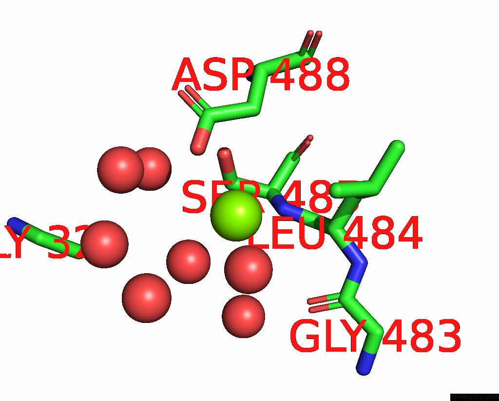
Mono view
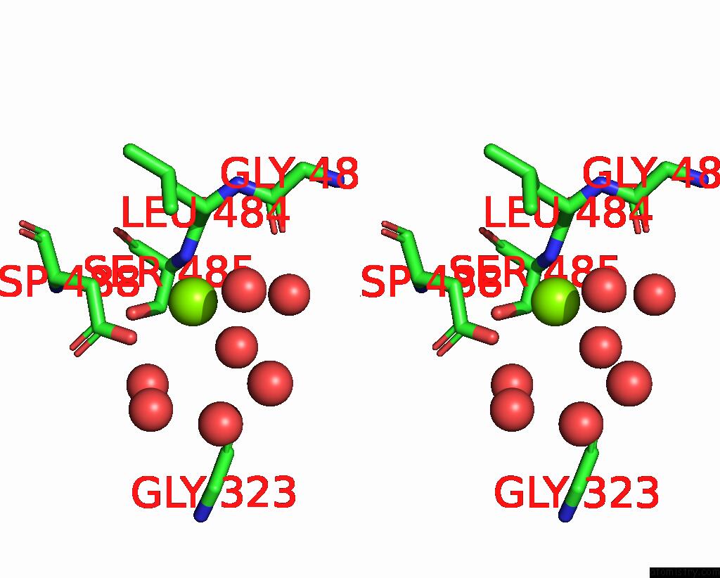
Stereo pair view
Mono view
Stereo pair view
A full contact list of Magnesium with other atoms in the Mg binding
site number 2 of Thioredoxin Glutathione Reductase of Schistosoma Mansoni at 1.25A Resolution. within 5.0Å range:
|
Magnesium binding site 3 out of 3 in 8pdd
Go back to
Magnesium binding site 3 out
of 3 in the Thioredoxin Glutathione Reductase of Schistosoma Mansoni at 1.25A Resolution.
Mono view
Stereo pair view
Mono view
Stereo pair view
A full contact list of Magnesium with other atoms in the Mg binding
site number 3 of Thioredoxin Glutathione Reductase of Schistosoma Mansoni at 1.25A Resolution. within 5.0Å range:
|
Reference:
L.R.De Souza Neto,
B.O.Montoya,
J.Brandao-Neto,
A.Verma,
S.Bowyer,
J.T.Moreira-Filho,
R.F.Dantas,
B.J.Neves,
C.H.Andrade,
F.Von Delft,
R.J.Owens,
N.Furnham,
F.P.Silva-Jr.
Fragment Library Screening By X-Ray Crystallography and Binding Site Analysis on Thioredoxin Glutathione Reductase of Schistosoma Mansoni. Sci Rep V. 14 1582 2024.
ISSN: ESSN 2045-2322
PubMed: 38238498
DOI: 10.1038/S41598-024-52018-2
Page generated: Fri Oct 4 15:59:54 2024
ISSN: ESSN 2045-2322
PubMed: 38238498
DOI: 10.1038/S41598-024-52018-2
Last articles
Zn in 9MJ5Zn in 9HNW
Zn in 9G0L
Zn in 9FNE
Zn in 9DZN
Zn in 9E0I
Zn in 9D32
Zn in 9DAK
Zn in 8ZXC
Zn in 8ZUF