Magnesium in PDB 8spc: Crystal Structure of the Cytochrome P450 Enzyme Rufo
Protein crystallography data
The structure of Crystal Structure of the Cytochrome P450 Enzyme Rufo, PDB code: 8spc
was solved by
B.D.Dratch,
K.M.Davis,
with X-Ray Crystallography technique. A brief refinement statistics is given in the table below:
| Resolution Low / High (Å) | 39.34 / 1.87 |
| Space group | P 41 21 2 |
| Cell size a, b, c (Å), α, β, γ (°) | 77.627, 77.627, 136.898, 90, 90, 90 |
| R / Rfree (%) | 19.3 / 21.7 |
Other elements in 8spc:
The structure of Crystal Structure of the Cytochrome P450 Enzyme Rufo also contains other interesting chemical elements:
| Chlorine | (Cl) | 2 atoms |
| Iron | (Fe) | 1 atom |
Magnesium Binding Sites:
The binding sites of Magnesium atom in the Crystal Structure of the Cytochrome P450 Enzyme Rufo
(pdb code 8spc). This binding sites where shown within
5.0 Angstroms radius around Magnesium atom.
In total only one binding site of Magnesium was determined in the Crystal Structure of the Cytochrome P450 Enzyme Rufo, PDB code: 8spc:
In total only one binding site of Magnesium was determined in the Crystal Structure of the Cytochrome P450 Enzyme Rufo, PDB code: 8spc:
Magnesium binding site 1 out of 1 in 8spc
Go back to
Magnesium binding site 1 out
of 1 in the Crystal Structure of the Cytochrome P450 Enzyme Rufo
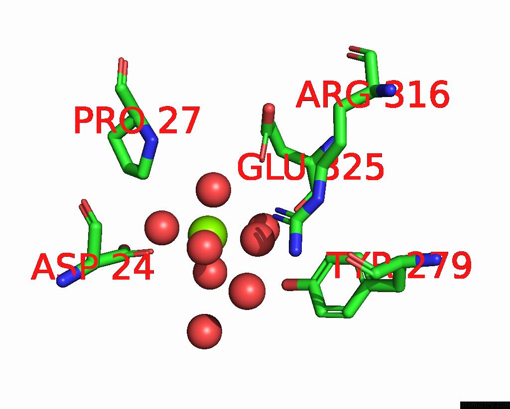
Mono view
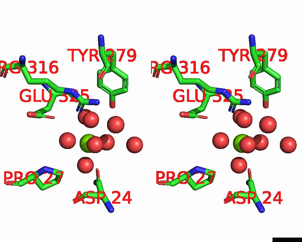
Stereo pair view
Mono view
Stereo pair view
A full contact list of Magnesium with other atoms in the Mg binding
site number 1 of Crystal Structure of the Cytochrome P450 Enzyme Rufo within 5.0Å range:
|
Reference:
B.D.Dratch,
K.L.Mcwhorter,
T.C.Blue,
S.K.Jones,
S.M.Horwitz,
K.M.Davis.
Insights Into Substrate Recognition By the Unusual Nitrating Enzyme Rufo. Acs Chem.Biol. V. 18 1713 2023.
ISSN: ESSN 1554-8937
PubMed: 37555759
DOI: 10.1021/ACSCHEMBIO.3C00328
Page generated: Fri Oct 4 19:55:53 2024
ISSN: ESSN 1554-8937
PubMed: 37555759
DOI: 10.1021/ACSCHEMBIO.3C00328
Last articles
Zn in 9MJ5Zn in 9HNW
Zn in 9G0L
Zn in 9FNE
Zn in 9DZN
Zn in 9E0I
Zn in 9D32
Zn in 9DAK
Zn in 8ZXC
Zn in 8ZUF