Magnesium in PDB 8t85: Structure of Rssb Bound to Beryllofluoride
Protein crystallography data
The structure of Structure of Rssb Bound to Beryllofluoride, PDB code: 8t85
was solved by
C.Brugger,
J.Schwartz,
A.M.Deaconescu,
with X-Ray Crystallography technique. A brief refinement statistics is given in the table below:
| Resolution Low / High (Å) | 67.99 / 2.38 |
| Space group | C 2 2 21 |
| Cell size a, b, c (Å), α, β, γ (°) | 41.366, 119.035, 135.973, 90, 90, 90 |
| R / Rfree (%) | 22.2 / 27.8 |
Other elements in 8t85:
The structure of Structure of Rssb Bound to Beryllofluoride also contains other interesting chemical elements:
| Fluorine | (F) | 3 atoms |
Magnesium Binding Sites:
The binding sites of Magnesium atom in the Structure of Rssb Bound to Beryllofluoride
(pdb code 8t85). This binding sites where shown within
5.0 Angstroms radius around Magnesium atom.
In total only one binding site of Magnesium was determined in the Structure of Rssb Bound to Beryllofluoride, PDB code: 8t85:
In total only one binding site of Magnesium was determined in the Structure of Rssb Bound to Beryllofluoride, PDB code: 8t85:
Magnesium binding site 1 out of 1 in 8t85
Go back to
Magnesium binding site 1 out
of 1 in the Structure of Rssb Bound to Beryllofluoride
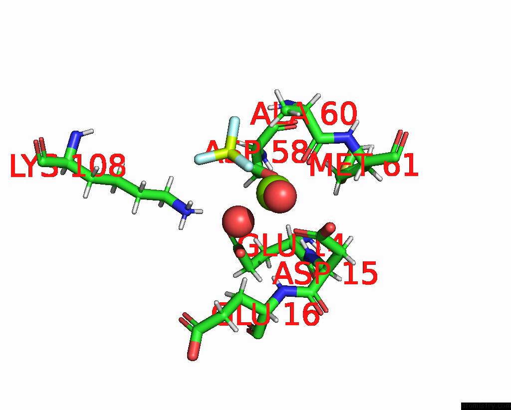
Mono view
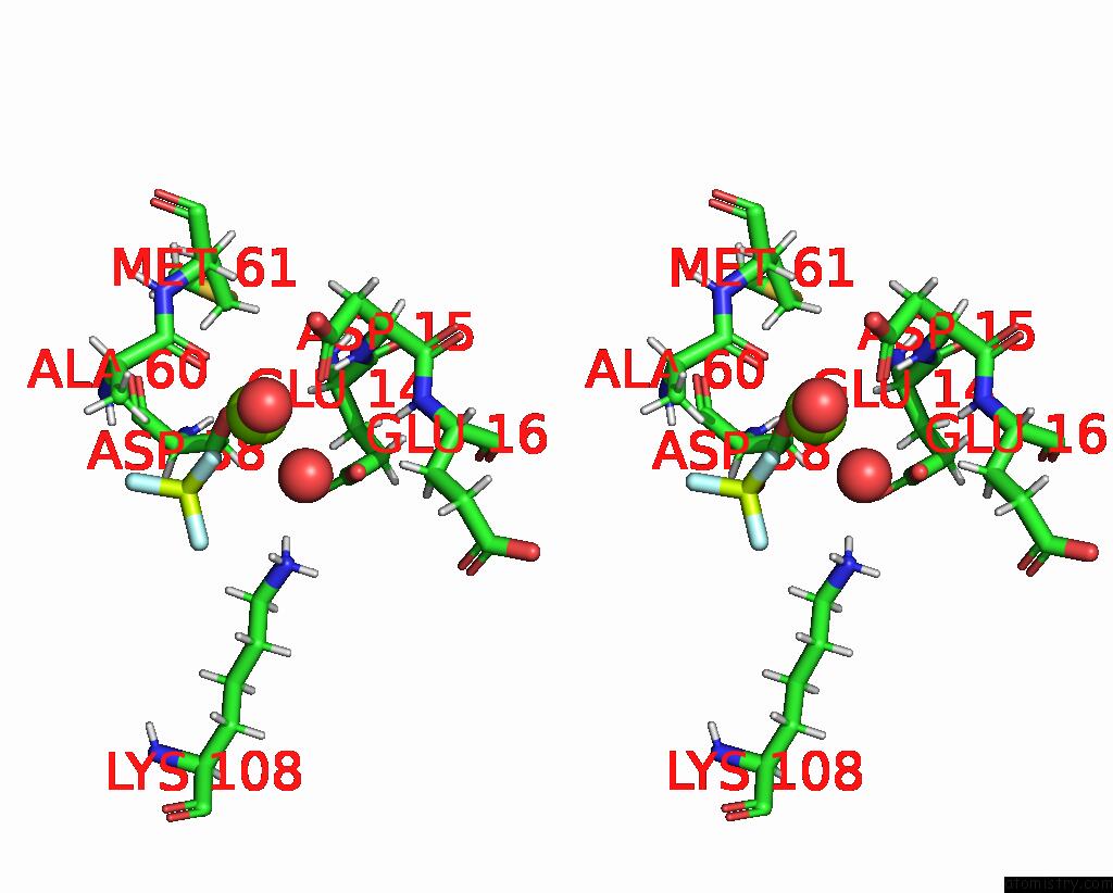
Stereo pair view
Mono view
Stereo pair view
A full contact list of Magnesium with other atoms in the Mg binding
site number 1 of Structure of Rssb Bound to Beryllofluoride within 5.0Å range:
|
Reference:
C.Brugger,
J.Schwartz,
S.Novick,
S.Tong,
J.R.Hoskins,
N.Majdalani,
R.Kim,
M.Filipovski,
S.Wickner,
S.Gottesman,
P.R.Griffin,
A.M.Deaconescu.
Structure of Phosphorylated-Like Rssb, the Adaptor Delivering Sigma S to the Clpxp Proteolytic Machinery, Reveals An Interface Switch For Activation. J.Biol.Chem. V. 299 05440 2023.
ISSN: ESSN 1083-351X
PubMed: 37949227
DOI: 10.1016/J.JBC.2023.105440
Page generated: Fri Oct 4 20:31:58 2024
ISSN: ESSN 1083-351X
PubMed: 37949227
DOI: 10.1016/J.JBC.2023.105440
Last articles
Zn in 9MJ5Zn in 9HNW
Zn in 9G0L
Zn in 9FNE
Zn in 9DZN
Zn in 9E0I
Zn in 9D32
Zn in 9DAK
Zn in 8ZXC
Zn in 8ZUF