Magnesium in PDB 8xdo: O-Methyltransferase From Lycoris Longituba Complexed with Mg and Sah
Protein crystallography data
The structure of O-Methyltransferase From Lycoris Longituba Complexed with Mg and Sah, PDB code: 8xdo
was solved by
Y.Y.H.Saw,
Y.Nakashima,
H.Morita,
with X-Ray Crystallography technique. A brief refinement statistics is given in the table below:
| Resolution Low / High (Å) | 47.73 / 1.37 |
| Space group | P 2 21 21 |
| Cell size a, b, c (Å), α, β, γ (°) | 49.637, 81.183, 118.027, 90, 90, 90 |
| R / Rfree (%) | 14.5 / 16.8 |
Magnesium Binding Sites:
The binding sites of Magnesium atom in the O-Methyltransferase From Lycoris Longituba Complexed with Mg and Sah
(pdb code 8xdo). This binding sites where shown within
5.0 Angstroms radius around Magnesium atom.
In total 2 binding sites of Magnesium where determined in the O-Methyltransferase From Lycoris Longituba Complexed with Mg and Sah, PDB code: 8xdo:
Jump to Magnesium binding site number: 1; 2;
In total 2 binding sites of Magnesium where determined in the O-Methyltransferase From Lycoris Longituba Complexed with Mg and Sah, PDB code: 8xdo:
Jump to Magnesium binding site number: 1; 2;
Magnesium binding site 1 out of 2 in 8xdo
Go back to
Magnesium binding site 1 out
of 2 in the O-Methyltransferase From Lycoris Longituba Complexed with Mg and Sah
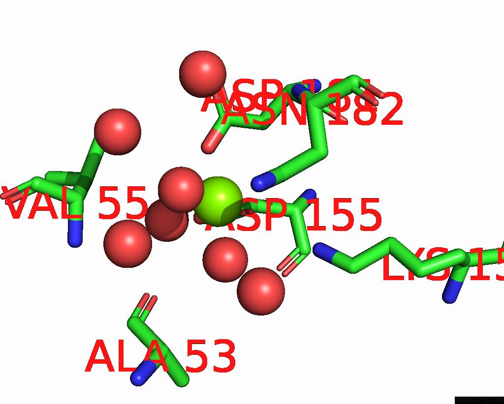
Mono view
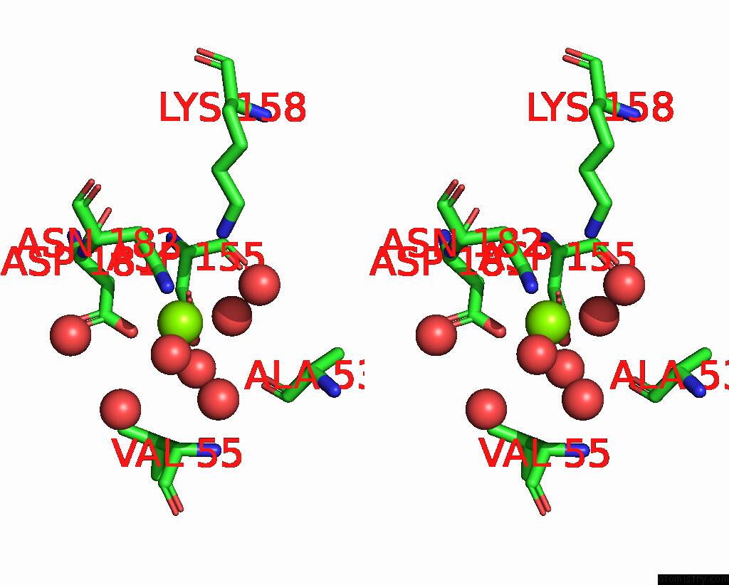
Stereo pair view
Mono view
Stereo pair view
A full contact list of Magnesium with other atoms in the Mg binding
site number 1 of O-Methyltransferase From Lycoris Longituba Complexed with Mg and Sah within 5.0Å range:
|
Magnesium binding site 2 out of 2 in 8xdo
Go back to
Magnesium binding site 2 out
of 2 in the O-Methyltransferase From Lycoris Longituba Complexed with Mg and Sah
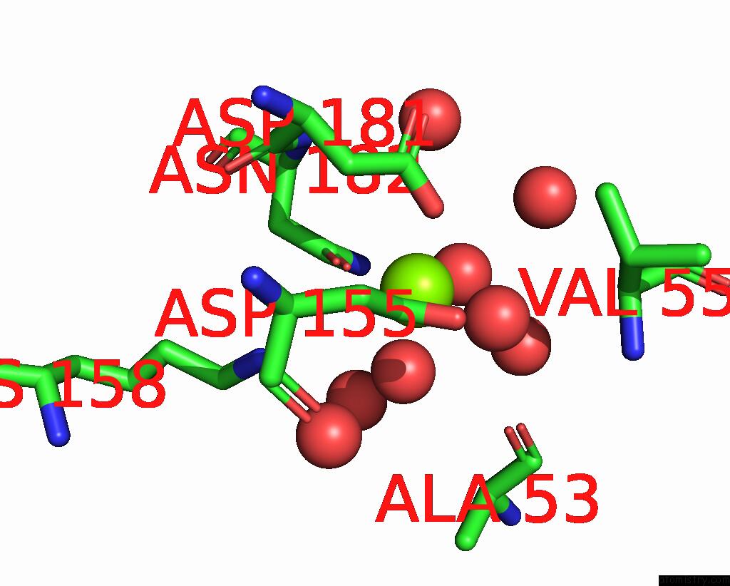
Mono view
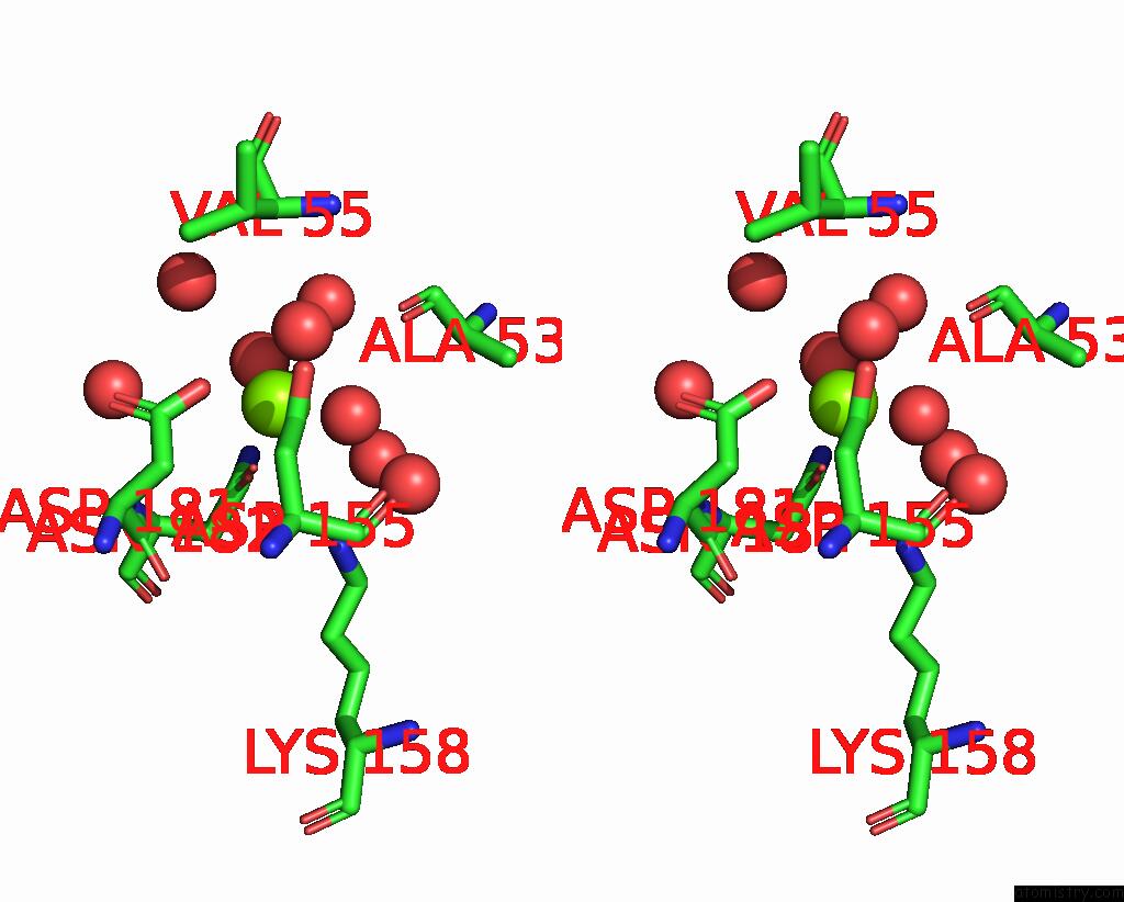
Stereo pair view
Mono view
Stereo pair view
A full contact list of Magnesium with other atoms in the Mg binding
site number 2 of O-Methyltransferase From Lycoris Longituba Complexed with Mg and Sah within 5.0Å range:
|
Reference:
S.Y.Y.Hnin,
Y.Nakashima,
T.Kodama,
H.Morita.
Structure-Based Catalytic Mechanism of Amaryllidaceae O-Methyltransferases Acs Catalysis 11865 2024.
ISSN: ESSN 2155-5435
DOI: 10.1021/ACSCATAL.4C03305
Page generated: Sat Oct 5 01:01:36 2024
ISSN: ESSN 2155-5435
DOI: 10.1021/ACSCATAL.4C03305
Last articles
Zn in 9MJ5Zn in 9HNW
Zn in 9G0L
Zn in 9FNE
Zn in 9DZN
Zn in 9E0I
Zn in 9D32
Zn in 9DAK
Zn in 8ZXC
Zn in 8ZUF