Magnesium in PDB 8xig: The Crystal Structure of the Aep Domain of Mpxv E5
Protein crystallography data
The structure of The Crystal Structure of the Aep Domain of Mpxv E5, PDB code: 8xig
was solved by
J.Gan,
W.Zhang,
with X-Ray Crystallography technique. A brief refinement statistics is given in the table below:
| Resolution Low / High (Å) | 27.41 / 1.65 |
| Space group | P 21 21 21 |
| Cell size a, b, c (Å), α, β, γ (°) | 51.982, 54.816, 74.845, 90, 90, 90 |
| R / Rfree (%) | 17.3 / 21.6 |
Magnesium Binding Sites:
The binding sites of Magnesium atom in the The Crystal Structure of the Aep Domain of Mpxv E5
(pdb code 8xig). This binding sites where shown within
5.0 Angstroms radius around Magnesium atom.
In total 2 binding sites of Magnesium where determined in the The Crystal Structure of the Aep Domain of Mpxv E5, PDB code: 8xig:
Jump to Magnesium binding site number: 1; 2;
In total 2 binding sites of Magnesium where determined in the The Crystal Structure of the Aep Domain of Mpxv E5, PDB code: 8xig:
Jump to Magnesium binding site number: 1; 2;
Magnesium binding site 1 out of 2 in 8xig
Go back to
Magnesium binding site 1 out
of 2 in the The Crystal Structure of the Aep Domain of Mpxv E5
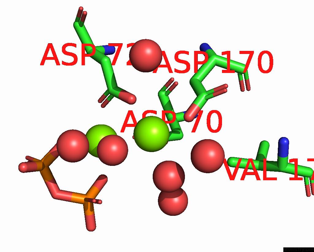
Mono view
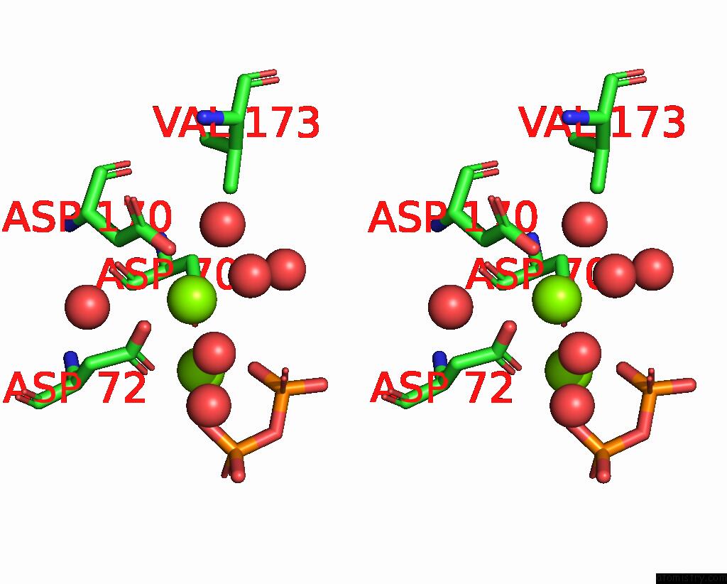
Stereo pair view
Mono view
Stereo pair view
A full contact list of Magnesium with other atoms in the Mg binding
site number 1 of The Crystal Structure of the Aep Domain of Mpxv E5 within 5.0Å range:
|
Magnesium binding site 2 out of 2 in 8xig
Go back to
Magnesium binding site 2 out
of 2 in the The Crystal Structure of the Aep Domain of Mpxv E5
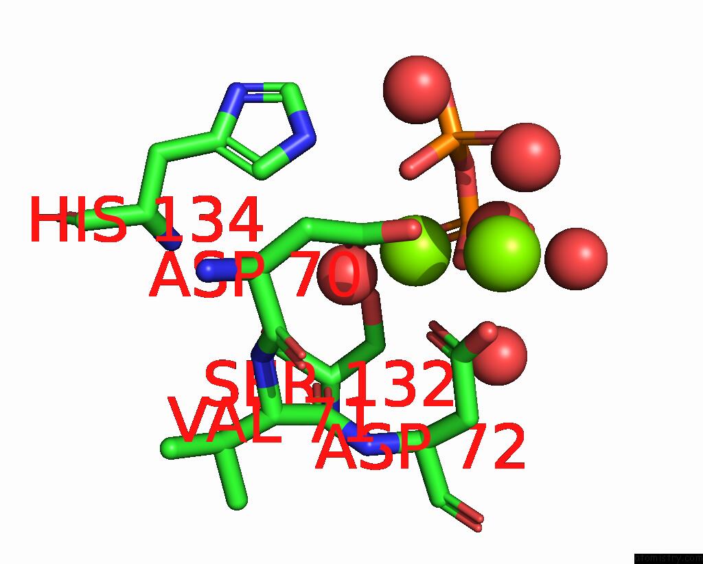
Mono view
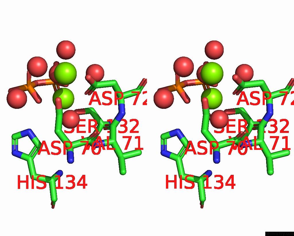
Stereo pair view
Mono view
Stereo pair view
A full contact list of Magnesium with other atoms in the Mg binding
site number 2 of The Crystal Structure of the Aep Domain of Mpxv E5 within 5.0Å range:
|
Reference:
J.Gan,
W.Zhang.
Structural and Functional Insights Into the Helicase Protein E5 of Mpox Virus. Cell Discov 2024.
ISSN: ESSN 2056-5968
Page generated: Sat Oct 5 01:05:05 2024
ISSN: ESSN 2056-5968
Last articles
Fe in 9CU0Fe in 9CJE
Fe in 9CJB
Fe in 9CJF
Fe in 9CTZ
Fe in 9CJD
Fe in 9CJC
Fe in 9CUF
Fe in 9DEU
Fe in 9CCB