Magnesium »
PDB 101d-1an0 »
109d »
Magnesium in PDB 109d: Variability in Dna Minor Groove Width Recognised By Ligand Binding: the Crystal Structure of A Bis-Benzimidazole Compound Bound to the Dna Duplex D(Cgcgaattcgcg)2
Protein crystallography data
The structure of Variability in Dna Minor Groove Width Recognised By Ligand Binding: the Crystal Structure of A Bis-Benzimidazole Compound Bound to the Dna Duplex D(Cgcgaattcgcg)2, PDB code: 109d
was solved by
A.Czarny,
D.W.Boykin,
A.A.Wood,
C.M.Nunn,
S.Neidle,
M.Zhao,
W.D.Wilson,
with X-Ray Crystallography technique. A brief refinement statistics is given in the table below:
| Resolution Low / High (Å) | 8.00 / 2.00 |
| Space group | P 21 21 21 |
| Cell size a, b, c (Å), α, β, γ (°) | 24.590, 40.440, 65.760, 90.00, 90.00, 90.00 |
| R / Rfree (%) | 19.7 / n/a |
Magnesium Binding Sites:
The binding sites of Magnesium atom in the Variability in Dna Minor Groove Width Recognised By Ligand Binding: the Crystal Structure of A Bis-Benzimidazole Compound Bound to the Dna Duplex D(Cgcgaattcgcg)2
(pdb code 109d). This binding sites where shown within
5.0 Angstroms radius around Magnesium atom.
In total only one binding site of Magnesium was determined in the Variability in Dna Minor Groove Width Recognised By Ligand Binding: the Crystal Structure of A Bis-Benzimidazole Compound Bound to the Dna Duplex D(Cgcgaattcgcg)2, PDB code: 109d:
In total only one binding site of Magnesium was determined in the Variability in Dna Minor Groove Width Recognised By Ligand Binding: the Crystal Structure of A Bis-Benzimidazole Compound Bound to the Dna Duplex D(Cgcgaattcgcg)2, PDB code: 109d:
Magnesium binding site 1 out of 1 in 109d
Go back to
Magnesium binding site 1 out
of 1 in the Variability in Dna Minor Groove Width Recognised By Ligand Binding: the Crystal Structure of A Bis-Benzimidazole Compound Bound to the Dna Duplex D(Cgcgaattcgcg)2
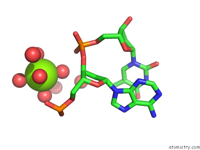
Mono view
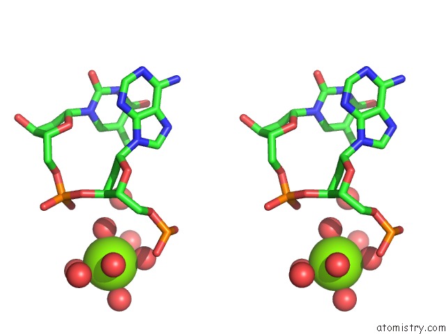
Stereo pair view
Mono view
Stereo pair view
A full contact list of Magnesium with other atoms in the Mg binding
site number 1 of Variability in Dna Minor Groove Width Recognised By Ligand Binding: the Crystal Structure of A Bis-Benzimidazole Compound Bound to the Dna Duplex D(Cgcgaattcgcg)2 within 5.0Å range:
|
Reference:
A.A.Wood,
C.M.Nunn,
A.Czarny,
D.W.Boykin,
S.Neidle.
Variability in Dna Minor Groove Width Recognised By Ligand Binding: the Crystal Structure of A Bis-Benzimidazole Compound Bound to the Dna Duplex D(Cgcgaattcgcg)2. Nucleic Acids Res. V. 23 3678 1995.
ISSN: ISSN 0305-1048
PubMed: 7478996
DOI: 10.1093/NAR/23.18.3678
Page generated: Tue Aug 13 01:59:46 2024
ISSN: ISSN 0305-1048
PubMed: 7478996
DOI: 10.1093/NAR/23.18.3678
Last articles
Ca in 5S9MCa in 5S9N
Ca in 5S9L
Ca in 5S8Q
Ca in 5S8P
Ca in 5S8O
Ca in 5S67
Ca in 5S65
Ca in 5S64
Ca in 5S63