Magnesium »
PDB 101d-1an0 »
1a05 »
Magnesium in PDB 1a05: Crystal Structure of the Complex of 3-Isopropylmalate Dehydrogenase From Thiobacillus Ferrooxidans with 3- Isopropylmalate
Enzymatic activity of Crystal Structure of the Complex of 3-Isopropylmalate Dehydrogenase From Thiobacillus Ferrooxidans with 3- Isopropylmalate
All present enzymatic activity of Crystal Structure of the Complex of 3-Isopropylmalate Dehydrogenase From Thiobacillus Ferrooxidans with 3- Isopropylmalate:
1.1.1.85;
1.1.1.85;
Protein crystallography data
The structure of Crystal Structure of the Complex of 3-Isopropylmalate Dehydrogenase From Thiobacillus Ferrooxidans with 3- Isopropylmalate, PDB code: 1a05
was solved by
K.Imada,
K.Inagaki,
H.Matsunami,
H.Kawaguchi,
H.Tanaka,
N.Tanaka,
K.Namba,
with X-Ray Crystallography technique. A brief refinement statistics is given in the table below:
| Resolution Low / High (Å) | 8.00 / 2.00 |
| Space group | P 21 21 2 |
| Cell size a, b, c (Å), α, β, γ (°) | 56.540, 114.240, 130.890, 90.00, 90.00, 90.00 |
| R / Rfree (%) | 19.8 / 27.5 |
Magnesium Binding Sites:
The binding sites of Magnesium atom in the Crystal Structure of the Complex of 3-Isopropylmalate Dehydrogenase From Thiobacillus Ferrooxidans with 3- Isopropylmalate
(pdb code 1a05). This binding sites where shown within
5.0 Angstroms radius around Magnesium atom.
In total 2 binding sites of Magnesium where determined in the Crystal Structure of the Complex of 3-Isopropylmalate Dehydrogenase From Thiobacillus Ferrooxidans with 3- Isopropylmalate, PDB code: 1a05:
Jump to Magnesium binding site number: 1; 2;
In total 2 binding sites of Magnesium where determined in the Crystal Structure of the Complex of 3-Isopropylmalate Dehydrogenase From Thiobacillus Ferrooxidans with 3- Isopropylmalate, PDB code: 1a05:
Jump to Magnesium binding site number: 1; 2;
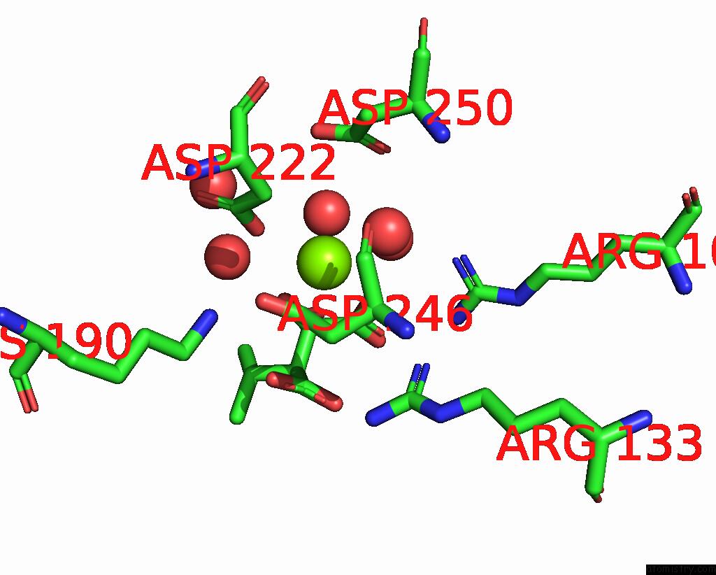
Magnesium binding site 1 out of 2 in 1a05
Go back to
Magnesium binding site 1 out
of 2 in the Crystal Structure of the Complex of 3-Isopropylmalate Dehydrogenase From Thiobacillus Ferrooxidans with 3- Isopropylmalate

Mono view
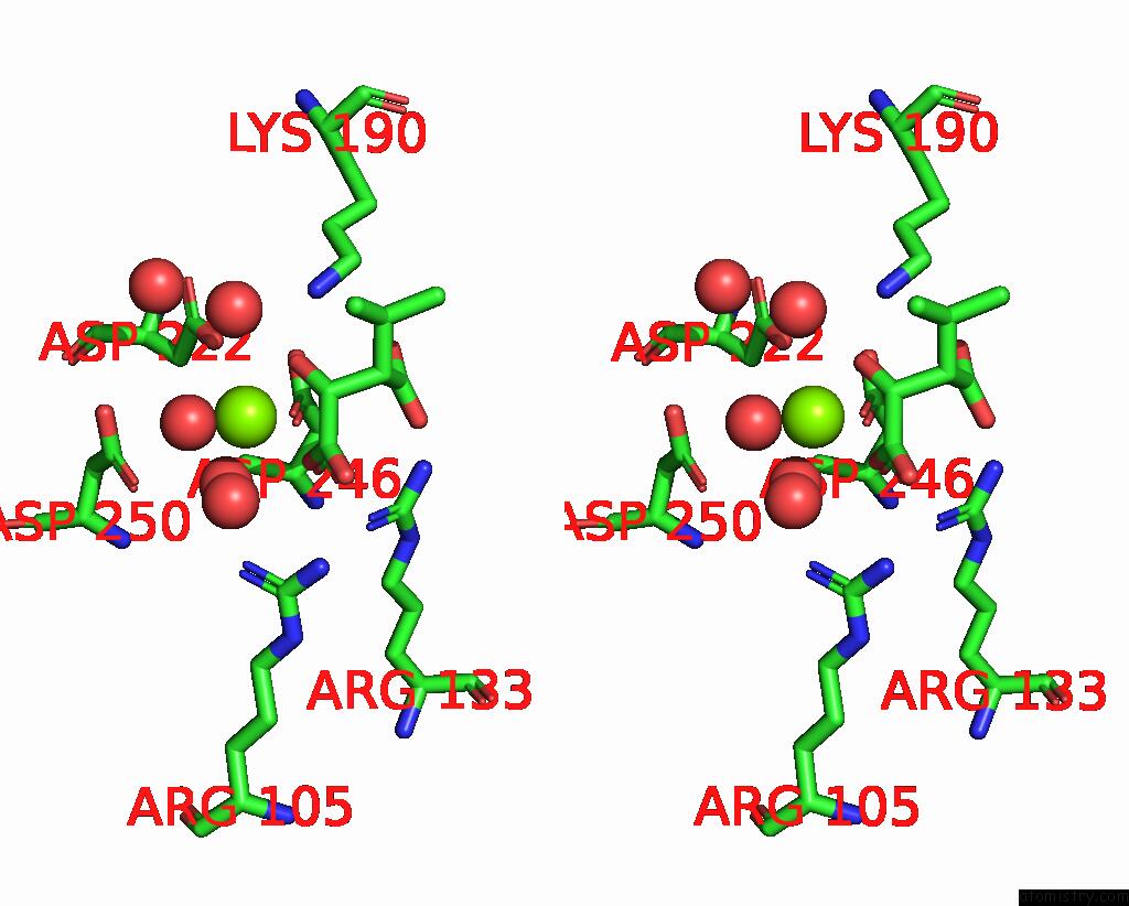
Stereo pair view
Mono view
Stereo pair view
A full contact list of Magnesium with other atoms in the Mg binding
site number 1 of Crystal Structure of the Complex of 3-Isopropylmalate Dehydrogenase From Thiobacillus Ferrooxidans with 3- Isopropylmalate within 5.0Å range:
|
Magnesium binding site 2 out of 2 in 1a05
Go back to
Magnesium binding site 2 out
of 2 in the Crystal Structure of the Complex of 3-Isopropylmalate Dehydrogenase From Thiobacillus Ferrooxidans with 3- Isopropylmalate
Mono view
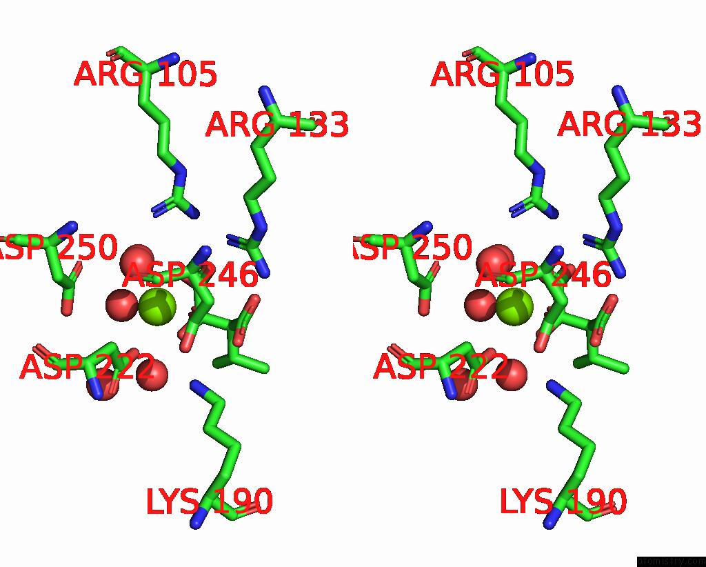
Stereo pair view
Mono view
Stereo pair view
A full contact list of Magnesium with other atoms in the Mg binding
site number 2 of Crystal Structure of the Complex of 3-Isopropylmalate Dehydrogenase From Thiobacillus Ferrooxidans with 3- Isopropylmalate within 5.0Å range:
|
Reference:
K.Imada,
K.Inagaki,
H.Matsunami,
H.Kawaguchi,
H.Tanaka,
N.Tanaka,
K.Namba.
Structure of 3-Isopropylmalate Dehydrogenase in Complex with 3-Isopropylmalate at 2.0 A Resolution: the Role of GLU88 in the Unique Substrate-Recognition Mechanism. Structure V. 6 971 1998.
ISSN: ISSN 0969-2126
PubMed: 9739088
DOI: 10.1016/S0969-2126(98)00099-9
Page generated: Tue Aug 13 02:00:08 2024
ISSN: ISSN 0969-2126
PubMed: 9739088
DOI: 10.1016/S0969-2126(98)00099-9
Last articles
Zn in 9MJ5Zn in 9HNW
Zn in 9G0L
Zn in 9FNE
Zn in 9DZN
Zn in 9E0I
Zn in 9D32
Zn in 9DAK
Zn in 8ZXC
Zn in 8ZUF