Magnesium »
PDB 101d-1an0 »
1a4r »
Magnesium in PDB 1a4r: G12V Mutant of Human Placental CDC42 Gtpase in the Gdp Form
Protein crystallography data
The structure of G12V Mutant of Human Placental CDC42 Gtpase in the Gdp Form, PDB code: 1a4r
was solved by
M.G.Rudolph,
I.R.Vetter,
A.Wittinghofer,
with X-Ray Crystallography technique. A brief refinement statistics is given in the table below:
| Resolution Low / High (Å) | 30.00 / 2.50 |
| Space group | P 41 21 2 |
| Cell size a, b, c (Å), α, β, γ (°) | 98.650, 98.650, 104.160, 90.00, 90.00, 90.00 |
| R / Rfree (%) | 22.8 / 28 |
Magnesium Binding Sites:
The binding sites of Magnesium atom in the G12V Mutant of Human Placental CDC42 Gtpase in the Gdp Form
(pdb code 1a4r). This binding sites where shown within
5.0 Angstroms radius around Magnesium atom.
In total only one binding site of Magnesium was determined in the G12V Mutant of Human Placental CDC42 Gtpase in the Gdp Form, PDB code: 1a4r:
In total only one binding site of Magnesium was determined in the G12V Mutant of Human Placental CDC42 Gtpase in the Gdp Form, PDB code: 1a4r:
Magnesium binding site 1 out of 1 in 1a4r
Go back to
Magnesium binding site 1 out
of 1 in the G12V Mutant of Human Placental CDC42 Gtpase in the Gdp Form
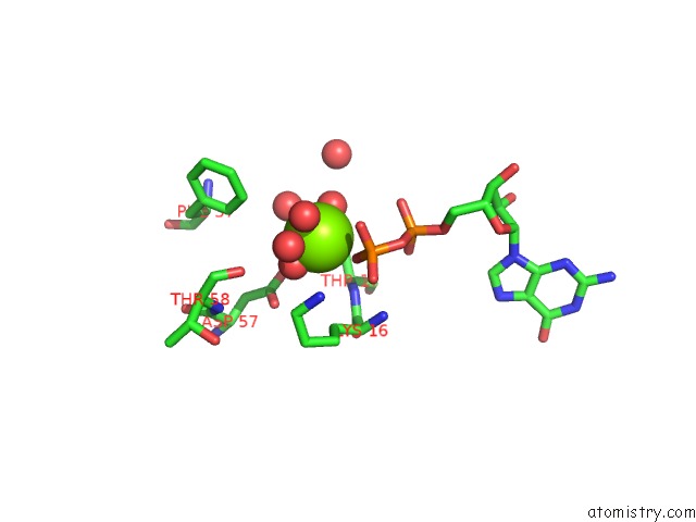
Mono view
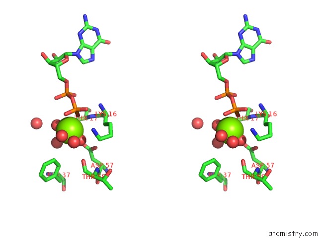
Stereo pair view
Mono view
Stereo pair view
A full contact list of Magnesium with other atoms in the Mg binding
site number 1 of G12V Mutant of Human Placental CDC42 Gtpase in the Gdp Form within 5.0Å range:
|
Reference:
M.G.Rudolph,
A.Wittinghofer,
I.R.Vetter.
Nucleotide Binding to the G12V-Mutant of CDC42 Investigated By X-Ray Diffraction and Fluorescence Spectroscopy: Two Different Nucleotide States in One Crystal. Protein Sci. V. 8 778 1999.
ISSN: ISSN 0961-8368
PubMed: 10211824
Page generated: Tue Aug 13 02:00:33 2024
ISSN: ISSN 0961-8368
PubMed: 10211824
Last articles
Cl in 7TP6Cl in 7TQ5
Cl in 7TQ4
Cl in 7TOJ
Cl in 7TP5
Cl in 7TO6
Cl in 7TNP
Cl in 7TO5
Cl in 7TN0
Cl in 7TNO