Magnesium »
PDB 1ao0-1bh2 »
1b4k »
Magnesium in PDB 1b4k: High Resolution Crystal Structure of A MG2-Dependent 5-Aminolevulinic Acid Dehydratase
Enzymatic activity of High Resolution Crystal Structure of A MG2-Dependent 5-Aminolevulinic Acid Dehydratase
All present enzymatic activity of High Resolution Crystal Structure of A MG2-Dependent 5-Aminolevulinic Acid Dehydratase:
4.2.1.24;
4.2.1.24;
Protein crystallography data
The structure of High Resolution Crystal Structure of A MG2-Dependent 5-Aminolevulinic Acid Dehydratase, PDB code: 1b4k
was solved by
N.Frankenberg,
D.Jahn,
D.W.Heinz,
with X-Ray Crystallography technique. A brief refinement statistics is given in the table below:
| Resolution Low / High (Å) | 51.30 / 1.67 |
| Space group | P 4 21 2 |
| Cell size a, b, c (Å), α, β, γ (°) | 128.240, 128.240, 86.220, 90.00, 90.00, 90.00 |
| R / Rfree (%) | 17.8 / 20.8 |
Magnesium Binding Sites:
The binding sites of Magnesium atom in the High Resolution Crystal Structure of A MG2-Dependent 5-Aminolevulinic Acid Dehydratase
(pdb code 1b4k). This binding sites where shown within
5.0 Angstroms radius around Magnesium atom.
In total only one binding site of Magnesium was determined in the High Resolution Crystal Structure of A MG2-Dependent 5-Aminolevulinic Acid Dehydratase, PDB code: 1b4k:
In total only one binding site of Magnesium was determined in the High Resolution Crystal Structure of A MG2-Dependent 5-Aminolevulinic Acid Dehydratase, PDB code: 1b4k:
Magnesium binding site 1 out of 1 in 1b4k
Go back to
Magnesium binding site 1 out
of 1 in the High Resolution Crystal Structure of A MG2-Dependent 5-Aminolevulinic Acid Dehydratase
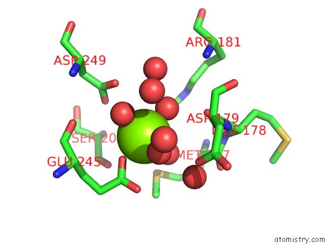
Mono view
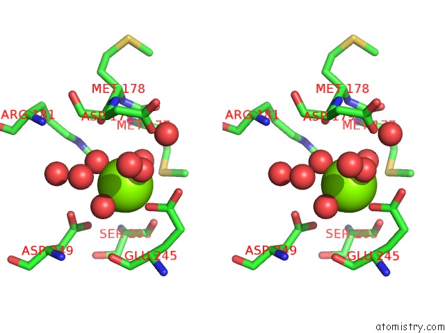
Stereo pair view
Mono view
Stereo pair view
A full contact list of Magnesium with other atoms in the Mg binding
site number 1 of High Resolution Crystal Structure of A MG2-Dependent 5-Aminolevulinic Acid Dehydratase within 5.0Å range:
|
Reference:
N.Frankenberg,
P.T.Erskine,
J.B.Cooper,
P.M.Shoolingin-Jordan,
D.Jahn,
D.W.Heinz.
High Resolution Crystal Structure of A MG2+-Dependent Porphobilinogen Synthase. J.Mol.Biol. V. 289 591 1999.
ISSN: ISSN 0022-2836
PubMed: 10356331
DOI: 10.1006/JMBI.1999.2808
Page generated: Tue Aug 13 02:10:09 2024
ISSN: ISSN 0022-2836
PubMed: 10356331
DOI: 10.1006/JMBI.1999.2808
Last articles
Zn in 9J0NZn in 9J0O
Zn in 9J0P
Zn in 9FJX
Zn in 9EKB
Zn in 9C0F
Zn in 9CAH
Zn in 9CH0
Zn in 9CH3
Zn in 9CH1