Magnesium »
PDB 1ao0-1bh2 »
1b7y »
Magnesium in PDB 1b7y: Phenylalanyl Trna Synthetase Complexed with Phenylalaninyl-Adenylate
Enzymatic activity of Phenylalanyl Trna Synthetase Complexed with Phenylalaninyl-Adenylate
All present enzymatic activity of Phenylalanyl Trna Synthetase Complexed with Phenylalaninyl-Adenylate:
6.1.1.20;
6.1.1.20;
Protein crystallography data
The structure of Phenylalanyl Trna Synthetase Complexed with Phenylalaninyl-Adenylate, PDB code: 1b7y
was solved by
L.Reshetnikova,
N.Moor,
O.Lavrik,
D.G.Vassylyev,
with X-Ray Crystallography technique. A brief refinement statistics is given in the table below:
| Resolution Low / High (Å) | 50.00 / 2.50 |
| Space group | P 32 2 1 |
| Cell size a, b, c (Å), α, β, γ (°) | 174.500, 174.500, 140.200, 90.00, 90.00, 120.00 |
| R / Rfree (%) | 23 / 26.7 |
Magnesium Binding Sites:
The binding sites of Magnesium atom in the Phenylalanyl Trna Synthetase Complexed with Phenylalaninyl-Adenylate
(pdb code 1b7y). This binding sites where shown within
5.0 Angstroms radius around Magnesium atom.
In total only one binding site of Magnesium was determined in the Phenylalanyl Trna Synthetase Complexed with Phenylalaninyl-Adenylate, PDB code: 1b7y:
In total only one binding site of Magnesium was determined in the Phenylalanyl Trna Synthetase Complexed with Phenylalaninyl-Adenylate, PDB code: 1b7y:
Magnesium binding site 1 out of 1 in 1b7y
Go back to
Magnesium binding site 1 out
of 1 in the Phenylalanyl Trna Synthetase Complexed with Phenylalaninyl-Adenylate
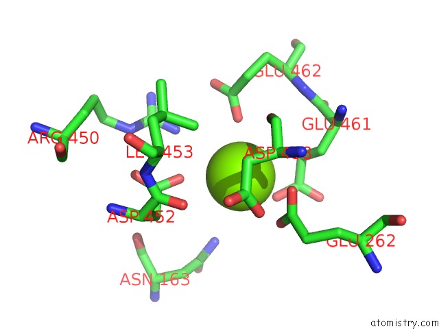
Mono view
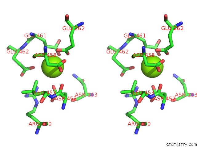
Stereo pair view
Mono view
Stereo pair view
A full contact list of Magnesium with other atoms in the Mg binding
site number 1 of Phenylalanyl Trna Synthetase Complexed with Phenylalaninyl-Adenylate within 5.0Å range:
|
Reference:
L.Reshetnikova,
N.Moor,
O.Lavrik,
D.G.Vassylyev.
Crystal Structures of Phenylalanyl-Trna Synthetase Complexed with Phenylalanine and A Phenylalanyl-Adenylate Analogue. J.Mol.Biol. V. 287 555 1999.
ISSN: ISSN 0022-2836
PubMed: 10092459
DOI: 10.1006/JMBI.1999.2617
Page generated: Sat Aug 9 20:10:58 2025
ISSN: ISSN 0022-2836
PubMed: 10092459
DOI: 10.1006/JMBI.1999.2617
Last articles
Mg in 6CA4Mg in 6C90
Mg in 6CA0
Mg in 6C9Y
Mg in 6C8Z
Mg in 6C8P
Mg in 6C8N
Mg in 6C8O
Mg in 6C8D
Mg in 6C8L