Magnesium »
PDB 1c5u-1cxz »
1c9k »
Magnesium in PDB 1c9k: The Three Dimensional Structure of Adenosylcobinamide Kinase/ Adenosylcobinamide Phosphate Gualylyltransferase (Cobu) Complexed with Gmp: Evidence For A Substrate Induced Transferase Active Site
Protein crystallography data
The structure of The Three Dimensional Structure of Adenosylcobinamide Kinase/ Adenosylcobinamide Phosphate Gualylyltransferase (Cobu) Complexed with Gmp: Evidence For A Substrate Induced Transferase Active Site, PDB code: 1c9k
was solved by
T.B.Thompson,
M.G.Thomas,
J.C.Esclante-Semerena,
I.Rayment,
with X-Ray Crystallography technique. A brief refinement statistics is given in the table below:
| Resolution Low / High (Å) | 30.00 / 2.20 |
| Space group | P 21 21 21 |
| Cell size a, b, c (Å), α, β, γ (°) | 58.380, 87.770, 101.640, 90.00, 90.00, 90.00 |
| R / Rfree (%) | n/a / n/a |
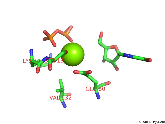
Magnesium Binding Sites:
The binding sites of Magnesium atom in the The Three Dimensional Structure of Adenosylcobinamide Kinase/ Adenosylcobinamide Phosphate Gualylyltransferase (Cobu) Complexed with Gmp: Evidence For A Substrate Induced Transferase Active Site
(pdb code 1c9k). This binding sites where shown within
5.0 Angstroms radius around Magnesium atom.
In total only one binding site of Magnesium was determined in the The Three Dimensional Structure of Adenosylcobinamide Kinase/ Adenosylcobinamide Phosphate Gualylyltransferase (Cobu) Complexed with Gmp: Evidence For A Substrate Induced Transferase Active Site, PDB code: 1c9k:
In total only one binding site of Magnesium was determined in the The Three Dimensional Structure of Adenosylcobinamide Kinase/ Adenosylcobinamide Phosphate Gualylyltransferase (Cobu) Complexed with Gmp: Evidence For A Substrate Induced Transferase Active Site, PDB code: 1c9k:
Magnesium binding site 1 out of 1 in 1c9k
Go back to
Magnesium binding site 1 out
of 1 in the The Three Dimensional Structure of Adenosylcobinamide Kinase/ Adenosylcobinamide Phosphate Gualylyltransferase (Cobu) Complexed with Gmp: Evidence For A Substrate Induced Transferase Active Site
Mono view

Stereo pair view
Mono view
Stereo pair view
A full contact list of Magnesium with other atoms in the Mg binding
site number 1 of The Three Dimensional Structure of Adenosylcobinamide Kinase/ Adenosylcobinamide Phosphate Gualylyltransferase (Cobu) Complexed with Gmp: Evidence For A Substrate Induced Transferase Active Site within 5.0Å range:
|
Reference:
T.B.Thompson,
M.G.Thomas,
J.C.Escalante-Semerena,
I.Rayment.
Three-Dimensional Structure of Adenosylcobinamide Kinase/Adenosylcobinamide Phosphate Guanylyltransferase (Cobu) Complexed with Gmp: Evidence For A Substrate-Induced Transferase Active Site. Biochemistry V. 38 12995 1999.
ISSN: ISSN 0006-2960
PubMed: 10529169
DOI: 10.1021/BI990910X
Page generated: Tue Aug 13 02:27:57 2024
ISSN: ISSN 0006-2960
PubMed: 10529169
DOI: 10.1021/BI990910X
Last articles
Fe in 8ZQDFe in 8ZEH
Fe in 8ZET
Fe in 8Z11
Fe in 9AZ2
Fe in 8ZK2
Fe in 9BCJ
Fe in 8ZXQ
Fe in 9AZ0
Fe in 9AZ1