Magnesium »
PDB 1c5u-1cxz »
1cg0 »
Magnesium in PDB 1cg0: Structure of Adenylosuccinate Synthetase From E. Coli Complexed with Hadacidin, Gdp, 6-Phosphoryl-Imp, and MG2+
Enzymatic activity of Structure of Adenylosuccinate Synthetase From E. Coli Complexed with Hadacidin, Gdp, 6-Phosphoryl-Imp, and MG2+
All present enzymatic activity of Structure of Adenylosuccinate Synthetase From E. Coli Complexed with Hadacidin, Gdp, 6-Phosphoryl-Imp, and MG2+:
6.3.4.4;
6.3.4.4;
Protein crystallography data
The structure of Structure of Adenylosuccinate Synthetase From E. Coli Complexed with Hadacidin, Gdp, 6-Phosphoryl-Imp, and MG2+, PDB code: 1cg0
was solved by
J.Y.Choe,
B.W.Poland,
H.Fromm,
R.Honzatko,
with X-Ray Crystallography technique. A brief refinement statistics is given in the table below:
| Resolution Low / High (Å) | 5.00 / 2.50 |
| Space group | P 32 2 1 |
| Cell size a, b, c (Å), α, β, γ (°) | 79.860, 79.860, 158.430, 90.00, 90.00, 120.00 |
| R / Rfree (%) | 16 / 25.9 |
Magnesium Binding Sites:
The binding sites of Magnesium atom in the Structure of Adenylosuccinate Synthetase From E. Coli Complexed with Hadacidin, Gdp, 6-Phosphoryl-Imp, and MG2+
(pdb code 1cg0). This binding sites where shown within
5.0 Angstroms radius around Magnesium atom.
In total only one binding site of Magnesium was determined in the Structure of Adenylosuccinate Synthetase From E. Coli Complexed with Hadacidin, Gdp, 6-Phosphoryl-Imp, and MG2+, PDB code: 1cg0:
In total only one binding site of Magnesium was determined in the Structure of Adenylosuccinate Synthetase From E. Coli Complexed with Hadacidin, Gdp, 6-Phosphoryl-Imp, and MG2+, PDB code: 1cg0:
Magnesium binding site 1 out of 1 in 1cg0
Go back to
Magnesium binding site 1 out
of 1 in the Structure of Adenylosuccinate Synthetase From E. Coli Complexed with Hadacidin, Gdp, 6-Phosphoryl-Imp, and MG2+
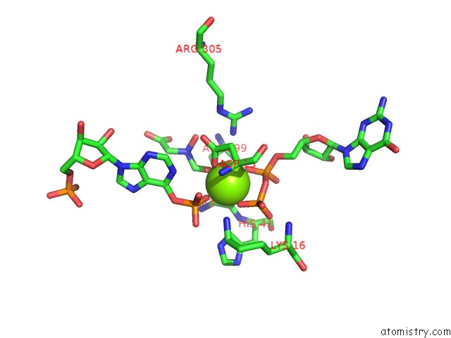
Mono view
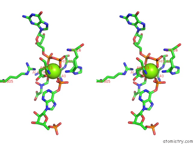
Stereo pair view
Mono view
Stereo pair view
A full contact list of Magnesium with other atoms in the Mg binding
site number 1 of Structure of Adenylosuccinate Synthetase From E. Coli Complexed with Hadacidin, Gdp, 6-Phosphoryl-Imp, and MG2+ within 5.0Å range:
|
Reference:
J.Y.Choe,
B.W.Poland,
H.J.Fromm,
R.B.Honzatko.
Mechanistic Implications From Crystalline Complexes of Wild-Type and Mutant Adenylosuccinate Synthetases From Escherichia Coli. Biochemistry V. 38 6953 1999.
ISSN: ISSN 0006-2960
PubMed: 10346917
DOI: 10.1021/BI990159S
Page generated: Tue Aug 13 02:28:22 2024
ISSN: ISSN 0006-2960
PubMed: 10346917
DOI: 10.1021/BI990159S
Last articles
F in 7H6WF in 7H5S
F in 7H5R
F in 7H5T
F in 7H5P
F in 7H5M
F in 7H5L
F in 7H5N
F in 7H5B
F in 7H57