Magnesium »
PDB 1c5u-1cxz »
1cw0 »
Magnesium in PDB 1cw0: Crystal Structure Analysis of Very Short Patch Repair (Vsr) Endonuclease in Complex with A Duplex Dna
Protein crystallography data
The structure of Crystal Structure Analysis of Very Short Patch Repair (Vsr) Endonuclease in Complex with A Duplex Dna, PDB code: 1cw0
was solved by
S.E.Tsutakawa,
H.Jingami,
K.Morikawa,
with X-Ray Crystallography technique. A brief refinement statistics is given in the table below:
| Resolution Low / High (Å) | 22.00 / 2.30 |
| Space group | P 65 |
| Cell size a, b, c (Å), α, β, γ (°) | 63.000, 63.000, 152.500, 90.00, 90.00, 120.00 |
| R / Rfree (%) | 20.4 / 23 |
Other elements in 1cw0:
The structure of Crystal Structure Analysis of Very Short Patch Repair (Vsr) Endonuclease in Complex with A Duplex Dna also contains other interesting chemical elements:
| Zinc | (Zn) | 1 atom |
Magnesium Binding Sites:
The binding sites of Magnesium atom in the Crystal Structure Analysis of Very Short Patch Repair (Vsr) Endonuclease in Complex with A Duplex Dna
(pdb code 1cw0). This binding sites where shown within
5.0 Angstroms radius around Magnesium atom.
In total 2 binding sites of Magnesium where determined in the Crystal Structure Analysis of Very Short Patch Repair (Vsr) Endonuclease in Complex with A Duplex Dna, PDB code: 1cw0:
Jump to Magnesium binding site number: 1; 2;
In total 2 binding sites of Magnesium where determined in the Crystal Structure Analysis of Very Short Patch Repair (Vsr) Endonuclease in Complex with A Duplex Dna, PDB code: 1cw0:
Jump to Magnesium binding site number: 1; 2;
Magnesium binding site 1 out of 2 in 1cw0
Go back to
Magnesium binding site 1 out
of 2 in the Crystal Structure Analysis of Very Short Patch Repair (Vsr) Endonuclease in Complex with A Duplex Dna
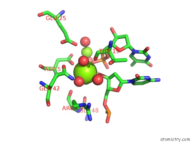
Mono view
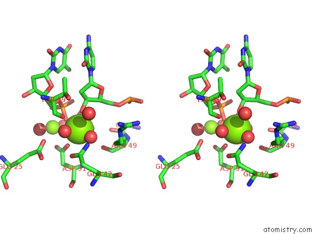
Stereo pair view
Mono view
Stereo pair view
A full contact list of Magnesium with other atoms in the Mg binding
site number 1 of Crystal Structure Analysis of Very Short Patch Repair (Vsr) Endonuclease in Complex with A Duplex Dna within 5.0Å range:
|
Magnesium binding site 2 out of 2 in 1cw0
Go back to
Magnesium binding site 2 out
of 2 in the Crystal Structure Analysis of Very Short Patch Repair (Vsr) Endonuclease in Complex with A Duplex Dna
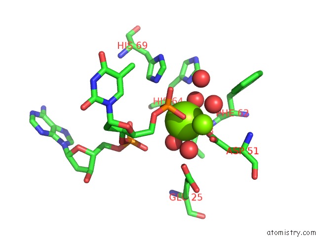
Mono view
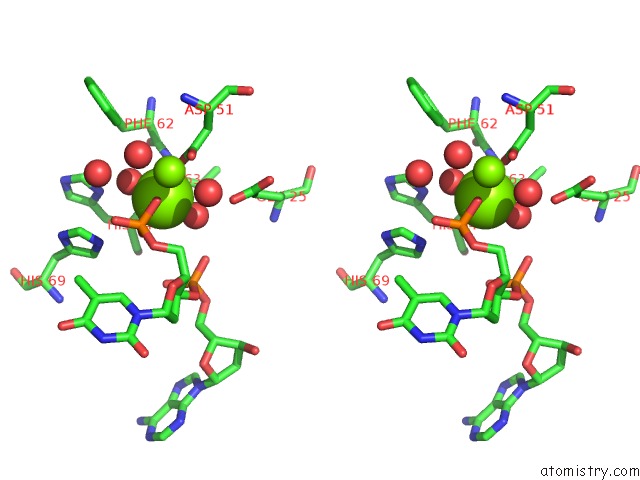
Stereo pair view
Mono view
Stereo pair view
A full contact list of Magnesium with other atoms in the Mg binding
site number 2 of Crystal Structure Analysis of Very Short Patch Repair (Vsr) Endonuclease in Complex with A Duplex Dna within 5.0Å range:
|
Reference:
S.E.Tsutakawa,
H.Jingami,
K.Morikawa.
Recognition of A Tg Mismatch: the Crystal Structure of Very Short Patch Repair Endonuclease in Complex with A Dna Duplex. Cell(Cambridge,Mass.) V. 99 615 1999.
ISSN: ISSN 0092-8674
PubMed: 10612397
DOI: 10.1016/S0092-8674(00)81550-0
Page generated: Sat Aug 9 20:25:54 2025
ISSN: ISSN 0092-8674
PubMed: 10612397
DOI: 10.1016/S0092-8674(00)81550-0
Last articles
Mg in 3W9TMg in 3WCW
Mg in 3WCA
Mg in 3WBH
Mg in 3W7W
Mg in 3WB2
Mg in 3WAG
Mg in 3W7F
Mg in 3WAD
Mg in 3W9S