Magnesium »
PDB 1cyq-1dak »
1d0z »
Magnesium in PDB 1d0z: Dictyostelium Myosin S1DC (Motor Domain Fragment) Complexed with P- Nitrophenyl Aminoethyldiphosphate Beryllium Trifluoride.
Protein crystallography data
The structure of Dictyostelium Myosin S1DC (Motor Domain Fragment) Complexed with P- Nitrophenyl Aminoethyldiphosphate Beryllium Trifluoride., PDB code: 1d0z
was solved by
A.M.Gulick,
C.B.Bauer,
J.B.Thoden,
E.Pate,
R.G.Yount,
I.Rayment,
with X-Ray Crystallography technique. A brief refinement statistics is given in the table below:
| Resolution Low / High (Å) | 25.00 / 2.00 |
| Space group | P 21 21 2 |
| Cell size a, b, c (Å), α, β, γ (°) | 103.987, 180.855, 54.101, 90.00, 90.00, 90.00 |
| R / Rfree (%) | n/a / n/a |
Other elements in 1d0z:
The structure of Dictyostelium Myosin S1DC (Motor Domain Fragment) Complexed with P- Nitrophenyl Aminoethyldiphosphate Beryllium Trifluoride. also contains other interesting chemical elements:
| Fluorine | (F) | 3 atoms |
Magnesium Binding Sites:
The binding sites of Magnesium atom in the Dictyostelium Myosin S1DC (Motor Domain Fragment) Complexed with P- Nitrophenyl Aminoethyldiphosphate Beryllium Trifluoride.
(pdb code 1d0z). This binding sites where shown within
5.0 Angstroms radius around Magnesium atom.
In total only one binding site of Magnesium was determined in the Dictyostelium Myosin S1DC (Motor Domain Fragment) Complexed with P- Nitrophenyl Aminoethyldiphosphate Beryllium Trifluoride., PDB code: 1d0z:
In total only one binding site of Magnesium was determined in the Dictyostelium Myosin S1DC (Motor Domain Fragment) Complexed with P- Nitrophenyl Aminoethyldiphosphate Beryllium Trifluoride., PDB code: 1d0z:
Magnesium binding site 1 out of 1 in 1d0z
Go back to
Magnesium binding site 1 out
of 1 in the Dictyostelium Myosin S1DC (Motor Domain Fragment) Complexed with P- Nitrophenyl Aminoethyldiphosphate Beryllium Trifluoride.
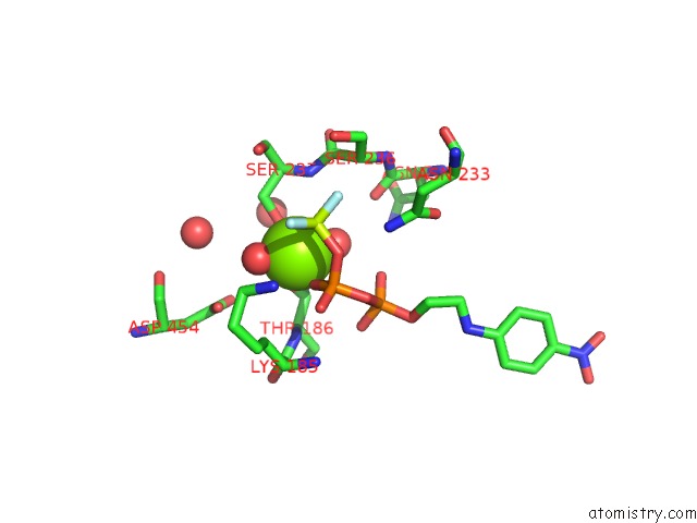
Mono view
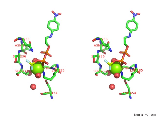
Stereo pair view
Mono view
Stereo pair view
A full contact list of Magnesium with other atoms in the Mg binding
site number 1 of Dictyostelium Myosin S1DC (Motor Domain Fragment) Complexed with P- Nitrophenyl Aminoethyldiphosphate Beryllium Trifluoride. within 5.0Å range:
|
Reference:
A.M.Gulick,
C.B.Bauer,
J.B.Thoden,
E.Pate,
R.G.Yount,
I.Rayment.
X-Ray Structures of the Dictyostelium Discoideum Myosin Motor Domain with Six Non-Nucleotide Analogs. J.Biol.Chem. V. 275 398 2000.
ISSN: ISSN 0021-9258
PubMed: 10617631
DOI: 10.1074/JBC.275.1.398
Page generated: Tue Aug 13 02:33:44 2024
ISSN: ISSN 0021-9258
PubMed: 10617631
DOI: 10.1074/JBC.275.1.398
Last articles
Zn in 9J0NZn in 9J0O
Zn in 9J0P
Zn in 9FJX
Zn in 9EKB
Zn in 9C0F
Zn in 9CAH
Zn in 9CH0
Zn in 9CH3
Zn in 9CH1