Magnesium »
PDB 1cyq-1dak »
1d38 »
Magnesium in PDB 1d38: Influence of Aglycone Modifications on the Binding of Anthracycline Drugs to Dna: the Molecular Structure of Idarubicin and 4-O-Demethyl-11-Deoxydoxorubicin Complexed to D(Cgatcg)
Protein crystallography data
The structure of Influence of Aglycone Modifications on the Binding of Anthracycline Drugs to Dna: the Molecular Structure of Idarubicin and 4-O-Demethyl-11-Deoxydoxorubicin Complexed to D(Cgatcg), PDB code: 1d38
was solved by
Y.-G.Gao,
A.H.-J.Wang,
with X-Ray Crystallography technique. A brief refinement statistics is given in the table below:
| Resolution Low / High (Å) | N/A / 1.70 |
| Space group | P 41 21 2 |
| Cell size a, b, c (Å), α, β, γ (°) | 28.130, 28.130, 53.680, 90.00, 90.00, 90.00 |
| R / Rfree (%) | n/a / n/a |
Magnesium Binding Sites:
The binding sites of Magnesium atom in the Influence of Aglycone Modifications on the Binding of Anthracycline Drugs to Dna: the Molecular Structure of Idarubicin and 4-O-Demethyl-11-Deoxydoxorubicin Complexed to D(Cgatcg)
(pdb code 1d38). This binding sites where shown within
5.0 Angstroms radius around Magnesium atom.
In total only one binding site of Magnesium was determined in the Influence of Aglycone Modifications on the Binding of Anthracycline Drugs to Dna: the Molecular Structure of Idarubicin and 4-O-Demethyl-11-Deoxydoxorubicin Complexed to D(Cgatcg), PDB code: 1d38:
In total only one binding site of Magnesium was determined in the Influence of Aglycone Modifications on the Binding of Anthracycline Drugs to Dna: the Molecular Structure of Idarubicin and 4-O-Demethyl-11-Deoxydoxorubicin Complexed to D(Cgatcg), PDB code: 1d38:
Magnesium binding site 1 out of 1 in 1d38
Go back to
Magnesium binding site 1 out
of 1 in the Influence of Aglycone Modifications on the Binding of Anthracycline Drugs to Dna: the Molecular Structure of Idarubicin and 4-O-Demethyl-11-Deoxydoxorubicin Complexed to D(Cgatcg)

Mono view
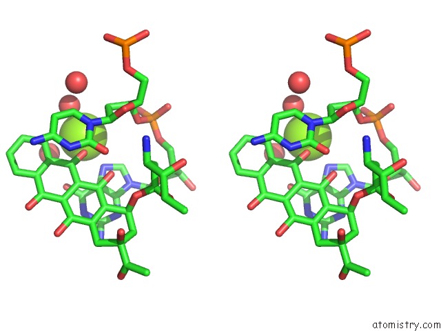
Stereo pair view
Mono view
Stereo pair view
A full contact list of Magnesium with other atoms in the Mg binding
site number 1 of Influence of Aglycone Modifications on the Binding of Anthracycline Drugs to Dna: the Molecular Structure of Idarubicin and 4-O-Demethyl-11-Deoxydoxorubicin Complexed to D(Cgatcg) within 5.0Å range:
|
Reference:
Y.G.Gao,
A.H.Wang.
Influence of Aglycone Modifications on the Binding of Anthracycline Drugs to Dna: the Molecular Structure of Idarubicin and 4-O-Demethyl-11-Deoxydoxorubicin Complexed to D(Cgatcg). Anti-Cancer Drug Des. V. 6 137 1991.
ISSN: ISSN 0266-9536
PubMed: 1872945
Page generated: Tue Aug 13 02:35:02 2024
ISSN: ISSN 0266-9536
PubMed: 1872945
Last articles
Zn in 9J0NZn in 9J0O
Zn in 9J0P
Zn in 9FJX
Zn in 9EKB
Zn in 9C0F
Zn in 9CAH
Zn in 9CH0
Zn in 9CH3
Zn in 9CH1