Magnesium »
PDB 1dam-1dtw »
1dbr »
Magnesium in PDB 1dbr: Hypoxanthine Guanine Xanthine
Protein crystallography data
The structure of Hypoxanthine Guanine Xanthine, PDB code: 1dbr
was solved by
M.A.Schumacher,
D.Carter,
D.Roos,
B.Ullman,
R.G.Brennan,
with X-Ray Crystallography technique. A brief refinement statistics is given in the table below:
| Resolution Low / High (Å) | 10.00 / 2.40 |
| Space group | P 1 21 1 |
| Cell size a, b, c (Å), α, β, γ (°) | 65.150, 109.220, 79.740, 90.00, 111.80, 90.00 |
| R / Rfree (%) | n/a / n/a |
Magnesium Binding Sites:
The binding sites of Magnesium atom in the Hypoxanthine Guanine Xanthine
(pdb code 1dbr). This binding sites where shown within
5.0 Angstroms radius around Magnesium atom.
In total 4 binding sites of Magnesium where determined in the Hypoxanthine Guanine Xanthine, PDB code: 1dbr:
Jump to Magnesium binding site number: 1; 2; 3; 4;
In total 4 binding sites of Magnesium where determined in the Hypoxanthine Guanine Xanthine, PDB code: 1dbr:
Jump to Magnesium binding site number: 1; 2; 3; 4;
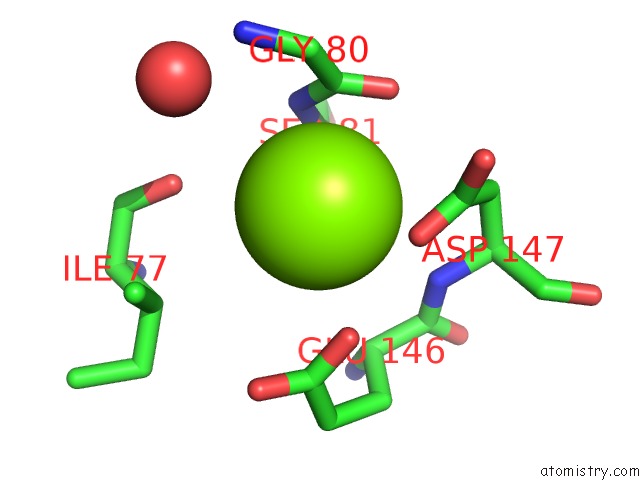
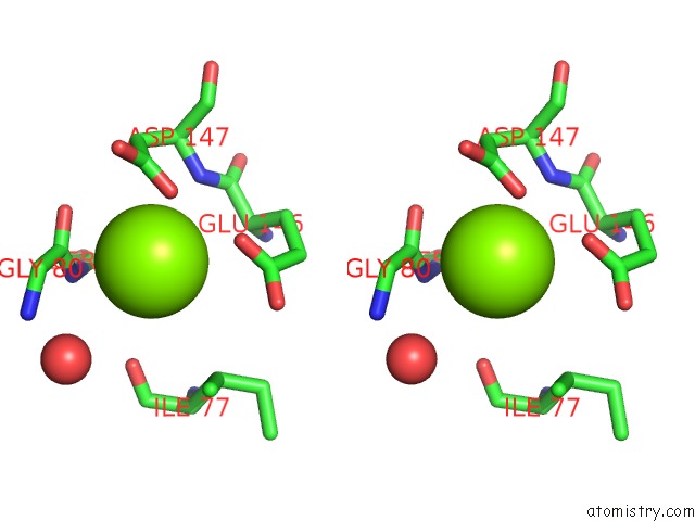
Magnesium binding site 1 out of 4 in 1dbr
Go back to
Magnesium binding site 1 out
of 4 in the Hypoxanthine Guanine Xanthine
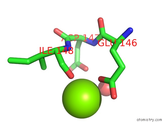
Mono view
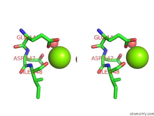
Stereo pair view
Mono view
Stereo pair view
A full contact list of Magnesium with other atoms in the Mg binding
site number 1 of Hypoxanthine Guanine Xanthine within 5.0Å range:
|
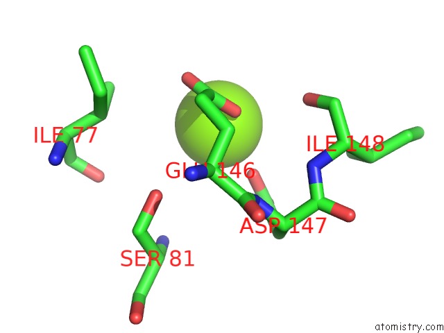
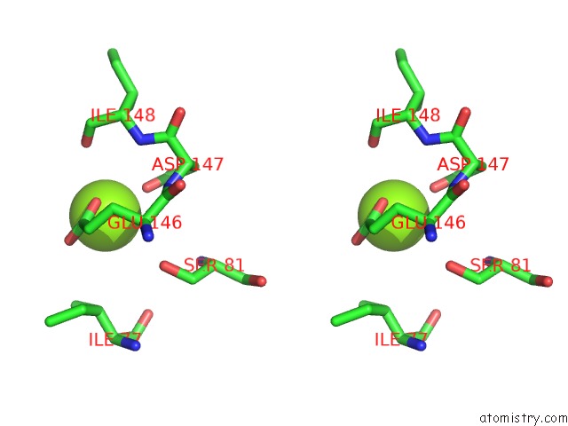
Magnesium binding site 2 out of 4 in 1dbr
Go back to
Magnesium binding site 2 out
of 4 in the Hypoxanthine Guanine Xanthine
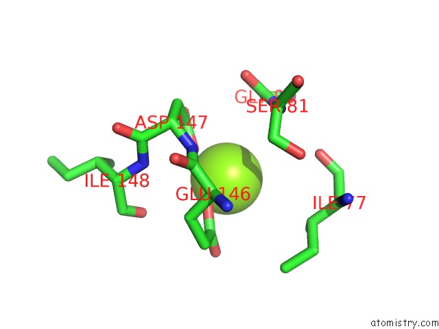
Mono view
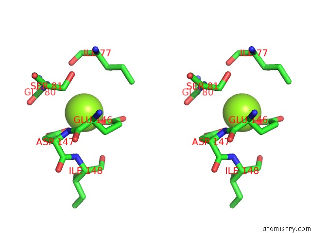
Stereo pair view
Mono view
Stereo pair view
A full contact list of Magnesium with other atoms in the Mg binding
site number 2 of Hypoxanthine Guanine Xanthine within 5.0Å range:
|
Magnesium binding site 3 out of 4 in 1dbr
Go back to
Magnesium binding site 3 out
of 4 in the Hypoxanthine Guanine Xanthine
Mono view
Stereo pair view
Mono view
Stereo pair view
A full contact list of Magnesium with other atoms in the Mg binding
site number 3 of Hypoxanthine Guanine Xanthine within 5.0Å range:
|
Magnesium binding site 4 out of 4 in 1dbr
Go back to
Magnesium binding site 4 out
of 4 in the Hypoxanthine Guanine Xanthine
Mono view
Stereo pair view
Mono view
Stereo pair view
A full contact list of Magnesium with other atoms in the Mg binding
site number 4 of Hypoxanthine Guanine Xanthine within 5.0Å range:
|
Reference:
M.A.Schumacher,
D.Carter,
D.S.Ross,
B.Ullman,
R.G.Brennan.
Crystal Structures of Toxoplasma Gondii Hgxprtase Reveal the Catalytic Role of A Long Flexible Loop. Nat.Struct.Biol. V. 3 881 1996.
ISSN: ISSN 1072-8368
PubMed: 8836106
DOI: 10.1038/NSB1096-881
Page generated: Tue Aug 13 02:37:45 2024
ISSN: ISSN 1072-8368
PubMed: 8836106
DOI: 10.1038/NSB1096-881
Last articles
Fe in 2YXOFe in 2YRS
Fe in 2YXC
Fe in 2YNM
Fe in 2YVJ
Fe in 2YP1
Fe in 2YU2
Fe in 2YU1
Fe in 2YQB
Fe in 2YOO