Magnesium »
PDB 1dam-1dtw »
1dfu »
Magnesium in PDB 1dfu: Crystal Structure of E.Coli Ribosomal Protein L25 Complexed with A 5S Rrna Fragment at 1.8 A Resolution
Protein crystallography data
The structure of Crystal Structure of E.Coli Ribosomal Protein L25 Complexed with A 5S Rrna Fragment at 1.8 A Resolution, PDB code: 1dfu
was solved by
M.Lu,
T.A.Steitz,
with X-Ray Crystallography technique. A brief refinement statistics is given in the table below:
| Resolution Low / High (Å) | 20.00 / 1.80 |
| Space group | C 2 2 21 |
| Cell size a, b, c (Å), α, β, γ (°) | 75.600, 76.600, 95.100, 90.00, 90.00, 90.00 |
| R / Rfree (%) | 20.7 / 22.5 |
Magnesium Binding Sites:
The binding sites of Magnesium atom in the Crystal Structure of E.Coli Ribosomal Protein L25 Complexed with A 5S Rrna Fragment at 1.8 A Resolution
(pdb code 1dfu). This binding sites where shown within
5.0 Angstroms radius around Magnesium atom.
In total 5 binding sites of Magnesium where determined in the Crystal Structure of E.Coli Ribosomal Protein L25 Complexed with A 5S Rrna Fragment at 1.8 A Resolution, PDB code: 1dfu:
Jump to Magnesium binding site number: 1; 2; 3; 4; 5;
In total 5 binding sites of Magnesium where determined in the Crystal Structure of E.Coli Ribosomal Protein L25 Complexed with A 5S Rrna Fragment at 1.8 A Resolution, PDB code: 1dfu:
Jump to Magnesium binding site number: 1; 2; 3; 4; 5;
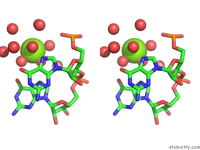
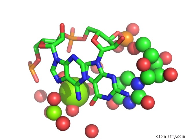
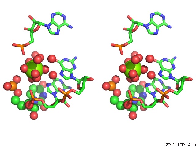
Magnesium binding site 1 out of 5 in 1dfu
Go back to
Magnesium binding site 1 out
of 5 in the Crystal Structure of E.Coli Ribosomal Protein L25 Complexed with A 5S Rrna Fragment at 1.8 A Resolution
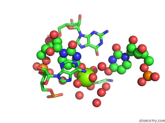
Mono view
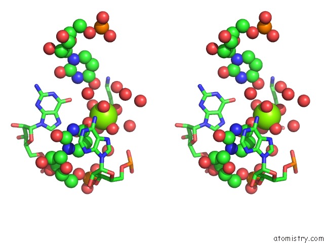
Stereo pair view
Mono view
Stereo pair view
A full contact list of Magnesium with other atoms in the Mg binding
site number 1 of Crystal Structure of E.Coli Ribosomal Protein L25 Complexed with A 5S Rrna Fragment at 1.8 A Resolution within 5.0Å range:
|
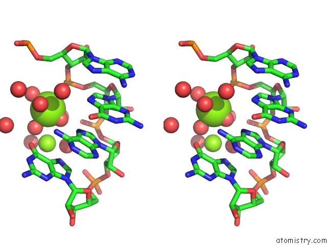
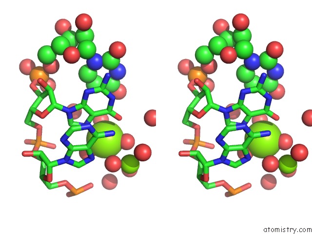
Magnesium binding site 2 out of 5 in 1dfu
Go back to
Magnesium binding site 2 out
of 5 in the Crystal Structure of E.Coli Ribosomal Protein L25 Complexed with A 5S Rrna Fragment at 1.8 A Resolution

Mono view
Stereo pair view
Mono view
Stereo pair view
A full contact list of Magnesium with other atoms in the Mg binding
site number 2 of Crystal Structure of E.Coli Ribosomal Protein L25 Complexed with A 5S Rrna Fragment at 1.8 A Resolution within 5.0Å range:
|
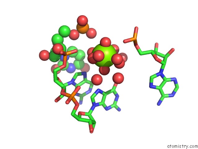
Magnesium binding site 3 out of 5 in 1dfu
Go back to
Magnesium binding site 3 out
of 5 in the Crystal Structure of E.Coli Ribosomal Protein L25 Complexed with A 5S Rrna Fragment at 1.8 A Resolution
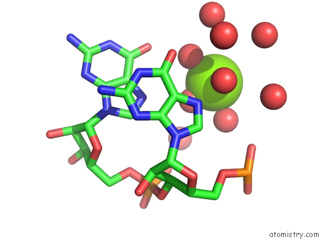
Mono view
Stereo pair view
Mono view
Stereo pair view
A full contact list of Magnesium with other atoms in the Mg binding
site number 3 of Crystal Structure of E.Coli Ribosomal Protein L25 Complexed with A 5S Rrna Fragment at 1.8 A Resolution within 5.0Å range:
|
Magnesium binding site 4 out of 5 in 1dfu
Go back to
Magnesium binding site 4 out
of 5 in the Crystal Structure of E.Coli Ribosomal Protein L25 Complexed with A 5S Rrna Fragment at 1.8 A Resolution
Mono view
Stereo pair view
Mono view
Stereo pair view
A full contact list of Magnesium with other atoms in the Mg binding
site number 4 of Crystal Structure of E.Coli Ribosomal Protein L25 Complexed with A 5S Rrna Fragment at 1.8 A Resolution within 5.0Å range:
|
Magnesium binding site 5 out of 5 in 1dfu
Go back to
Magnesium binding site 5 out
of 5 in the Crystal Structure of E.Coli Ribosomal Protein L25 Complexed with A 5S Rrna Fragment at 1.8 A Resolution
Mono view
Stereo pair view
Mono view
Stereo pair view
A full contact list of Magnesium with other atoms in the Mg binding
site number 5 of Crystal Structure of E.Coli Ribosomal Protein L25 Complexed with A 5S Rrna Fragment at 1.8 A Resolution within 5.0Å range:
|
Reference:
M.Lu,
T.A.Steitz.
Structure of Escherichia Coli Ribosomal Protein L25 Complexed with A 5S Rrna Fragment at 1.8-A Resolution. Proc.Natl.Acad.Sci.Usa V. 97 2023 2000.
ISSN: ISSN 0027-8424
PubMed: 10696113
DOI: 10.1073/PNAS.97.5.2023
Page generated: Tue Aug 13 02:39:27 2024
ISSN: ISSN 0027-8424
PubMed: 10696113
DOI: 10.1073/PNAS.97.5.2023
Last articles
Zn in 9J0NZn in 9J0O
Zn in 9J0P
Zn in 9FJX
Zn in 9EKB
Zn in 9C0F
Zn in 9CAH
Zn in 9CH0
Zn in 9CH3
Zn in 9CH1