Magnesium »
PDB 1dam-1dtw »
1dqn »
Magnesium in PDB 1dqn: Crystal Structure of Giardia Guanine Phosphoribosyltransferase Complexed with A Transition State Analogue
Enzymatic activity of Crystal Structure of Giardia Guanine Phosphoribosyltransferase Complexed with A Transition State Analogue
All present enzymatic activity of Crystal Structure of Giardia Guanine Phosphoribosyltransferase Complexed with A Transition State Analogue:
2.4.2.8;
2.4.2.8;
Protein crystallography data
The structure of Crystal Structure of Giardia Guanine Phosphoribosyltransferase Complexed with A Transition State Analogue, PDB code: 1dqn
was solved by
W.Shi,
N.R.Munagala,
C.C.Wang,
C.M.Li,
P.C.Tyler,
R.H.Furneaux,
C.Grubmeyer,
V.L.Schramm,
S.C.Almo,
with X-Ray Crystallography technique. A brief refinement statistics is given in the table below:
| Resolution Low / High (Å) | 20.00 / 1.75 |
| Space group | P 21 21 21 |
| Cell size a, b, c (Å), α, β, γ (°) | 56.632, 71.553, 123.080, 90.00, 90.00, 90.00 |
| R / Rfree (%) | 20.4 / 23.4 |
Magnesium Binding Sites:
The binding sites of Magnesium atom in the Crystal Structure of Giardia Guanine Phosphoribosyltransferase Complexed with A Transition State Analogue
(pdb code 1dqn). This binding sites where shown within
5.0 Angstroms radius around Magnesium atom.
In total 2 binding sites of Magnesium where determined in the Crystal Structure of Giardia Guanine Phosphoribosyltransferase Complexed with A Transition State Analogue, PDB code: 1dqn:
Jump to Magnesium binding site number: 1; 2;
In total 2 binding sites of Magnesium where determined in the Crystal Structure of Giardia Guanine Phosphoribosyltransferase Complexed with A Transition State Analogue, PDB code: 1dqn:
Jump to Magnesium binding site number: 1; 2;
Magnesium binding site 1 out of 2 in 1dqn
Go back to
Magnesium binding site 1 out
of 2 in the Crystal Structure of Giardia Guanine Phosphoribosyltransferase Complexed with A Transition State Analogue
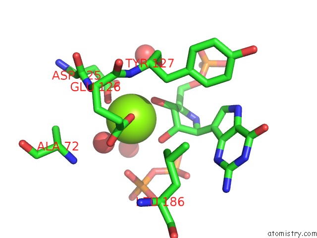
Mono view
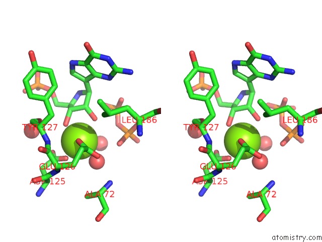
Stereo pair view
Mono view
Stereo pair view
A full contact list of Magnesium with other atoms in the Mg binding
site number 1 of Crystal Structure of Giardia Guanine Phosphoribosyltransferase Complexed with A Transition State Analogue within 5.0Å range:
|
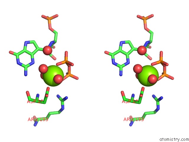
Magnesium binding site 2 out of 2 in 1dqn
Go back to
Magnesium binding site 2 out
of 2 in the Crystal Structure of Giardia Guanine Phosphoribosyltransferase Complexed with A Transition State Analogue
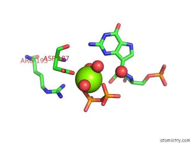
Mono view
Stereo pair view
Mono view
Stereo pair view
A full contact list of Magnesium with other atoms in the Mg binding
site number 2 of Crystal Structure of Giardia Guanine Phosphoribosyltransferase Complexed with A Transition State Analogue within 5.0Å range:
|
Reference:
W.Shi,
N.R.Munagala,
C.C.Wang,
C.M.Li,
P.C.Tyler,
R.H.Furneaux,
C.Grubmeyer,
V.L.Schramm,
S.C.Almo.
Crystal Structures of Giardia Lamblia Guanine Phosphoribosyltransferase at 1.75 A(,). Biochemistry V. 39 6781 2000.
ISSN: ISSN 0006-2960
PubMed: 10841757
DOI: 10.1021/BI000128T
Page generated: Tue Aug 13 02:42:15 2024
ISSN: ISSN 0006-2960
PubMed: 10841757
DOI: 10.1021/BI000128T
Last articles
Zn in 9J0NZn in 9J0O
Zn in 9J0P
Zn in 9FJX
Zn in 9EKB
Zn in 9C0F
Zn in 9CAH
Zn in 9CH0
Zn in 9CH3
Zn in 9CH1