Magnesium »
PDB 1dam-1dtw »
1dqv »
Magnesium in PDB 1dqv: Crystal Structure of Synaptotagmin III C2A/C2B
Protein crystallography data
The structure of Crystal Structure of Synaptotagmin III C2A/C2B, PDB code: 1dqv
was solved by
R.B.Sutton,
J.A.Ernst,
A.T.Brunger,
with X-Ray Crystallography technique. A brief refinement statistics is given in the table below:
| Resolution Low / High (Å) | 50.00 / 3.20 |
| Space group | P 62 2 2 |
| Cell size a, b, c (Å), α, β, γ (°) | 125.959, 125.959, 118.438, 90.00, 90.00, 120.00 |
| R / Rfree (%) | 29.3 / 34.8 |
Magnesium Binding Sites:
The binding sites of Magnesium atom in the Crystal Structure of Synaptotagmin III C2A/C2B
(pdb code 1dqv). This binding sites where shown within
5.0 Angstroms radius around Magnesium atom.
In total 4 binding sites of Magnesium where determined in the Crystal Structure of Synaptotagmin III C2A/C2B, PDB code: 1dqv:
Jump to Magnesium binding site number: 1; 2; 3; 4;
In total 4 binding sites of Magnesium where determined in the Crystal Structure of Synaptotagmin III C2A/C2B, PDB code: 1dqv:
Jump to Magnesium binding site number: 1; 2; 3; 4;
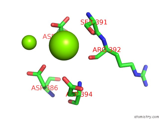
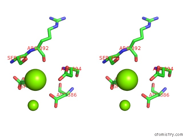
Magnesium binding site 1 out of 4 in 1dqv
Go back to
Magnesium binding site 1 out
of 4 in the Crystal Structure of Synaptotagmin III C2A/C2B
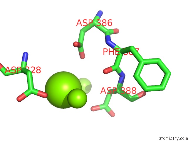
Mono view
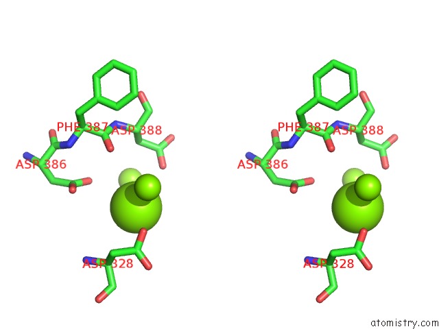
Stereo pair view
Mono view
Stereo pair view
A full contact list of Magnesium with other atoms in the Mg binding
site number 1 of Crystal Structure of Synaptotagmin III C2A/C2B within 5.0Å range:
|
Magnesium binding site 2 out of 4 in 1dqv
Go back to
Magnesium binding site 2 out
of 4 in the Crystal Structure of Synaptotagmin III C2A/C2B
Mono view
Stereo pair view
Mono view
Stereo pair view
A full contact list of Magnesium with other atoms in the Mg binding
site number 2 of Crystal Structure of Synaptotagmin III C2A/C2B within 5.0Å range:
|
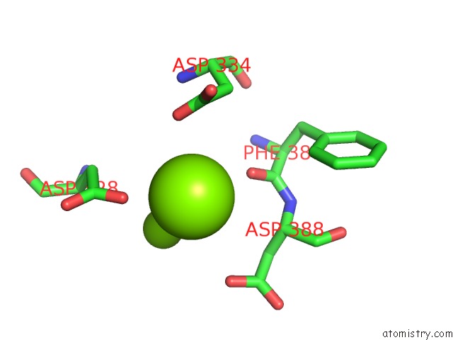
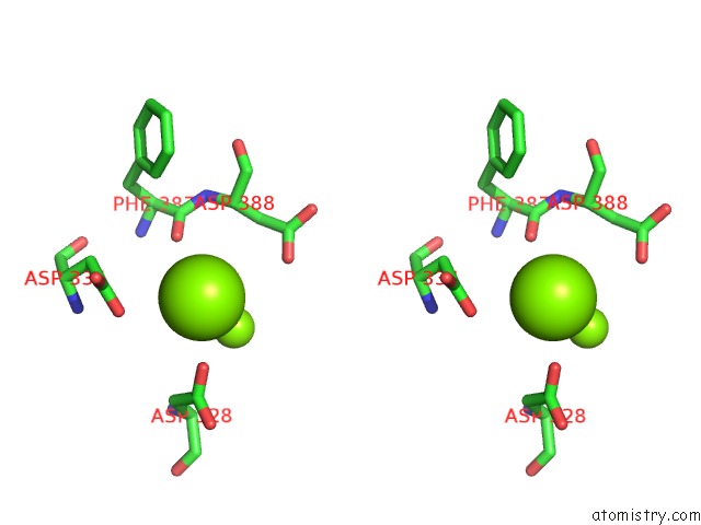
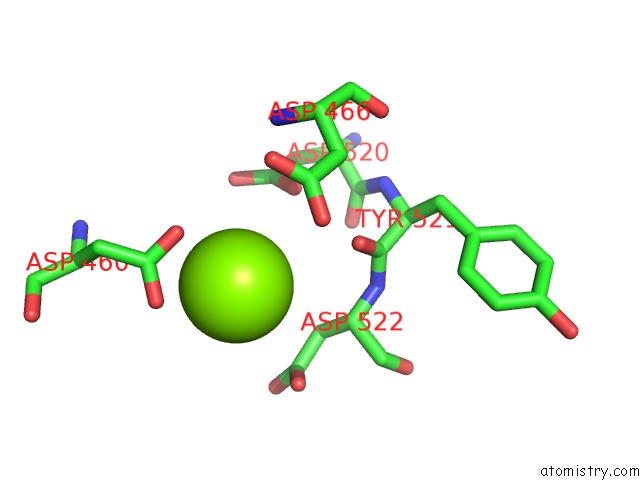
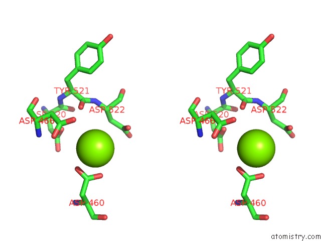
Magnesium binding site 3 out of 4 in 1dqv
Go back to
Magnesium binding site 3 out
of 4 in the Crystal Structure of Synaptotagmin III C2A/C2B
Mono view
Stereo pair view
Mono view
Stereo pair view
A full contact list of Magnesium with other atoms in the Mg binding
site number 3 of Crystal Structure of Synaptotagmin III C2A/C2B within 5.0Å range:
|
Magnesium binding site 4 out of 4 in 1dqv
Go back to
Magnesium binding site 4 out
of 4 in the Crystal Structure of Synaptotagmin III C2A/C2B
Mono view
Stereo pair view
Mono view
Stereo pair view
A full contact list of Magnesium with other atoms in the Mg binding
site number 4 of Crystal Structure of Synaptotagmin III C2A/C2B within 5.0Å range:
|
Reference:
R.B.Sutton,
J.A.Ernst,
A.T.Brunger.
Crystal Structure of the Cytosolic C2A-C2B Domains of Synaptotagmin III. Implications For Ca(+2)-Independent Snare Complex Interaction. J.Cell Biol. V. 147 589 1999.
ISSN: ISSN 0021-9525
PubMed: 10545502
DOI: 10.1083/JCB.147.3.589
Page generated: Tue Aug 13 02:42:46 2024
ISSN: ISSN 0021-9525
PubMed: 10545502
DOI: 10.1083/JCB.147.3.589
Last articles
Zn in 9MJ5Zn in 9HNW
Zn in 9G0L
Zn in 9FNE
Zn in 9DZN
Zn in 9E0I
Zn in 9D32
Zn in 9DAK
Zn in 8ZXC
Zn in 8ZUF