Magnesium »
PDB 1dam-1dtw »
1dtw »
Magnesium in PDB 1dtw: Human Branched-Chain Alpha-Keto Acid Dehydrogenase
Enzymatic activity of Human Branched-Chain Alpha-Keto Acid Dehydrogenase
All present enzymatic activity of Human Branched-Chain Alpha-Keto Acid Dehydrogenase:
1.2.4.4;
1.2.4.4;
Protein crystallography data
The structure of Human Branched-Chain Alpha-Keto Acid Dehydrogenase, PDB code: 1dtw
was solved by
A.Aevarsson,
J.L.Chuang,
R.M.Wynn,
S.Turley,
D.T.Chuang,
W.G.J.Hol,
with X-Ray Crystallography technique. A brief refinement statistics is given in the table below:
| Resolution Low / High (Å) | 50.00 / 2.70 |
| Space group | P 31 2 1 |
| Cell size a, b, c (Å), α, β, γ (°) | 143.760, 143.760, 69.160, 90.00, 90.00, 120.00 |
| R / Rfree (%) | 22.4 / 27.9 |
Other elements in 1dtw:
The structure of Human Branched-Chain Alpha-Keto Acid Dehydrogenase also contains other interesting chemical elements:
| Potassium | (K) | 2 atoms |
Magnesium Binding Sites:
The binding sites of Magnesium atom in the Human Branched-Chain Alpha-Keto Acid Dehydrogenase
(pdb code 1dtw). This binding sites where shown within
5.0 Angstroms radius around Magnesium atom.
In total only one binding site of Magnesium was determined in the Human Branched-Chain Alpha-Keto Acid Dehydrogenase, PDB code: 1dtw:
In total only one binding site of Magnesium was determined in the Human Branched-Chain Alpha-Keto Acid Dehydrogenase, PDB code: 1dtw:
Magnesium binding site 1 out of 1 in 1dtw
Go back to
Magnesium binding site 1 out
of 1 in the Human Branched-Chain Alpha-Keto Acid Dehydrogenase
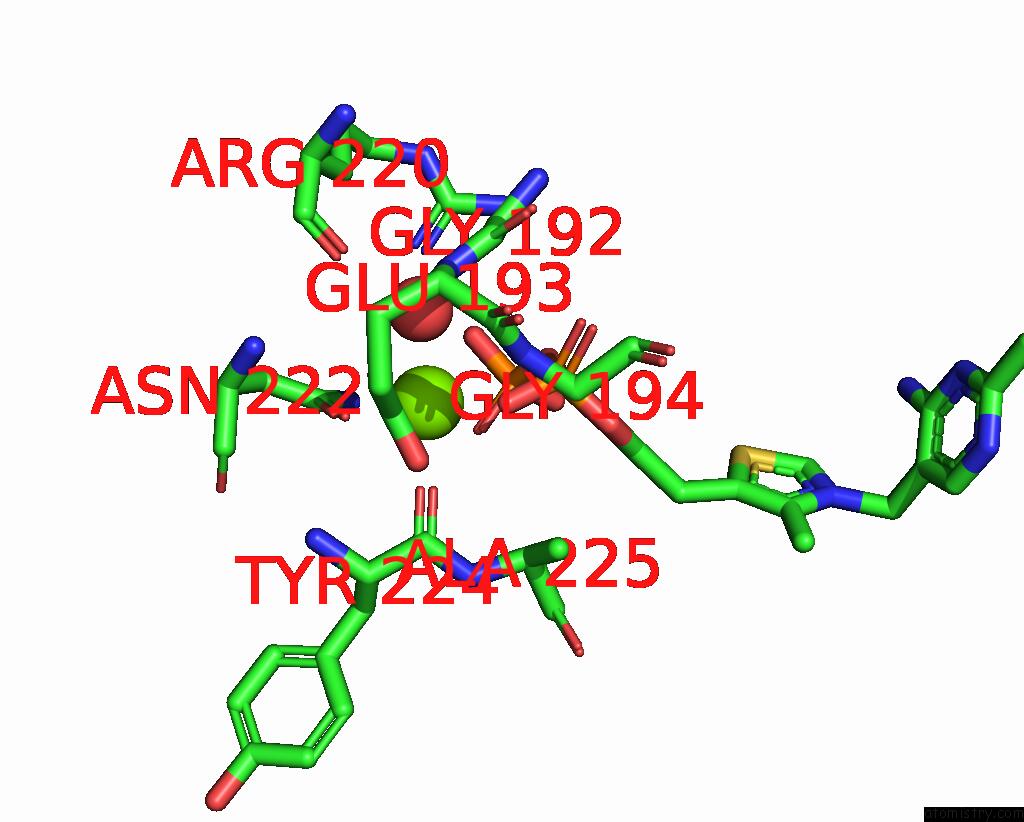
Mono view
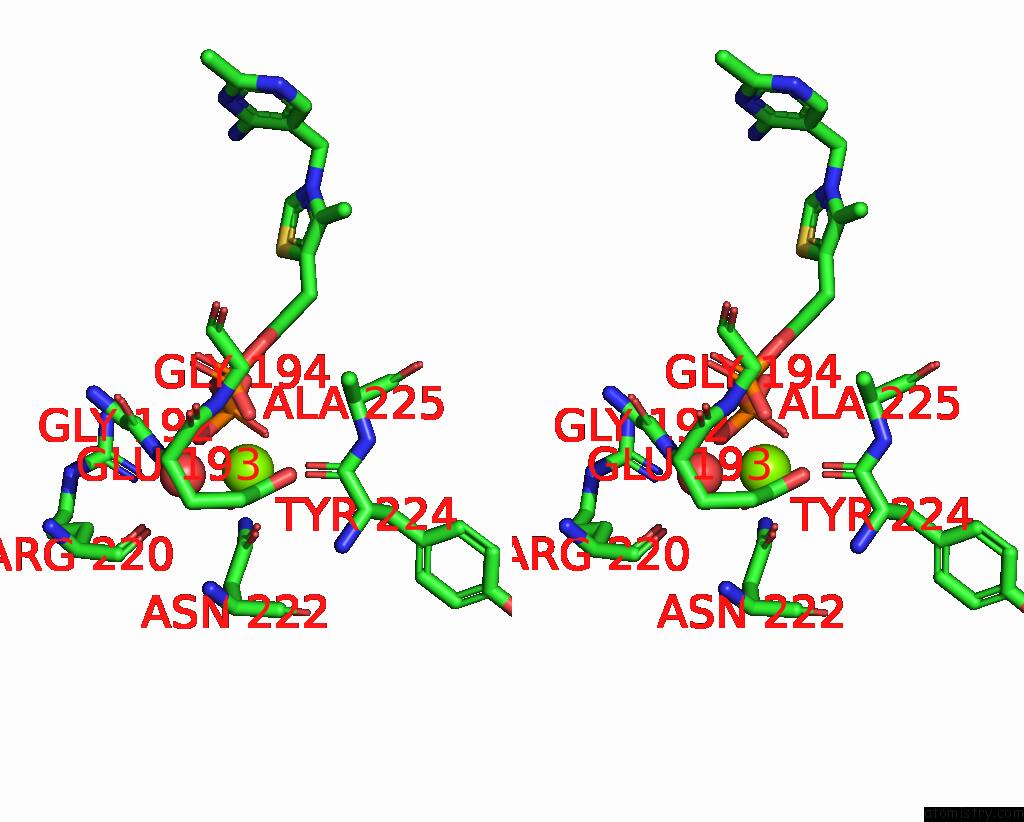
Stereo pair view
Mono view
Stereo pair view
A full contact list of Magnesium with other atoms in the Mg binding
site number 1 of Human Branched-Chain Alpha-Keto Acid Dehydrogenase within 5.0Å range:
|
Reference:
A.Aevarsson,
J.L.Chuang,
R.M.Wynn,
S.Turley,
D.T.Chuang,
W.G.Hol.
Crystal Structure of Human Branched-Chain Alpha-Ketoacid Dehydrogenase and the Molecular Basis of Multienzyme Complex Deficiency in Maple Syrup Urine Disease. Structure Fold.Des. V. 8 277 2000.
ISSN: ISSN 0969-2126
PubMed: 10745006
DOI: 10.1016/S0969-2126(00)00105-2
Page generated: Tue Aug 13 02:43:54 2024
ISSN: ISSN 0969-2126
PubMed: 10745006
DOI: 10.1016/S0969-2126(00)00105-2
Last articles
Zn in 9J0NZn in 9J0O
Zn in 9J0P
Zn in 9FJX
Zn in 9EKB
Zn in 9C0F
Zn in 9CAH
Zn in 9CH0
Zn in 9CH3
Zn in 9CH1