Magnesium »
PDB 1duh-1e9a »
1duh »
Magnesium in PDB 1duh: Crystal Structure of the Conserved Domain IV of E. Coli 4.5S Rna
Protein crystallography data
The structure of Crystal Structure of the Conserved Domain IV of E. Coli 4.5S Rna, PDB code: 1duh
was solved by
L.Jovine,
T.Hainzl,
C.Oubridge,
W.G.Scott,
J.Li,
T.K.Sixma,
A.Wonacott,
T.Skarzynski,
K.Nagai,
with X-Ray Crystallography technique. A brief refinement statistics is given in the table below:
| Resolution Low / High (Å) | 22.50 / 2.70 |
| Space group | P 32 2 1 |
| Cell size a, b, c (Å), α, β, γ (°) | 69.697, 69.697, 84.102, 90.00, 90.00, 120.00 |
| R / Rfree (%) | 23 / 24.5 |
Other elements in 1duh:
The structure of Crystal Structure of the Conserved Domain IV of E. Coli 4.5S Rna also contains other interesting chemical elements:
| Lutetium | (Lu) | 1 atom |
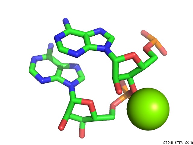
Magnesium Binding Sites:
The binding sites of Magnesium atom in the Crystal Structure of the Conserved Domain IV of E. Coli 4.5S Rna
(pdb code 1duh). This binding sites where shown within
5.0 Angstroms radius around Magnesium atom.
In total only one binding site of Magnesium was determined in the Crystal Structure of the Conserved Domain IV of E. Coli 4.5S Rna, PDB code: 1duh:
In total only one binding site of Magnesium was determined in the Crystal Structure of the Conserved Domain IV of E. Coli 4.5S Rna, PDB code: 1duh:
Magnesium binding site 1 out of 1 in 1duh
Go back to
Magnesium binding site 1 out
of 1 in the Crystal Structure of the Conserved Domain IV of E. Coli 4.5S Rna
Mono view
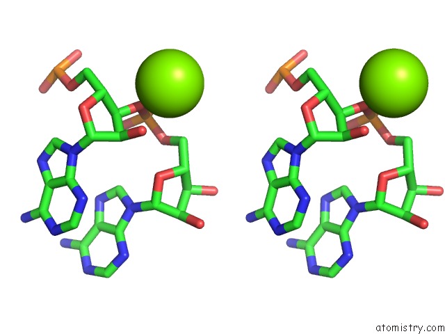
Stereo pair view
Mono view
Stereo pair view
A full contact list of Magnesium with other atoms in the Mg binding
site number 1 of Crystal Structure of the Conserved Domain IV of E. Coli 4.5S Rna within 5.0Å range:
|
Reference:
L.Jovine,
T.Hainzl,
C.Oubridge,
W.G.Scott,
J.Li,
T.K.Sixma,
A.Wonacott,
T.Skarzynski,
K.Nagai.
Crystal Structure of the Ffh and Ef-G Binding Sites in the Conserved Domain IV of Escherichia Coli 4.5S Rna. Structure Fold.Des. V. 8 527 2000.
ISSN: ISSN 0969-2126
PubMed: 10801497
DOI: 10.1016/S0969-2126(00)00137-4
Page generated: Tue Aug 13 02:45:51 2024
ISSN: ISSN 0969-2126
PubMed: 10801497
DOI: 10.1016/S0969-2126(00)00137-4
Last articles
Fe in 2YXOFe in 2YRS
Fe in 2YXC
Fe in 2YNM
Fe in 2YVJ
Fe in 2YP1
Fe in 2YU2
Fe in 2YU1
Fe in 2YQB
Fe in 2YOO