Magnesium »
PDB 1duh-1e9a »
1dul »
Magnesium in PDB 1dul: Structure of the Ribonucleoprotein Core of the E. Coli Signal Recognition Particle
Protein crystallography data
The structure of Structure of the Ribonucleoprotein Core of the E. Coli Signal Recognition Particle, PDB code: 1dul
was solved by
R.T.Batey,
R.P.Rambo,
L.Lucast,
B.Rha,
J.Doudna,
with X-Ray Crystallography technique. A brief refinement statistics is given in the table below:
| Resolution Low / High (Å) | 28.83 / 1.80 |
| Space group | C 1 2 1 |
| Cell size a, b, c (Å), α, β, γ (°) | 136.567, 78.291, 32.849, 90.00, 96.14, 90.00 |
| R / Rfree (%) | 19.9 / 22.1 |
Other elements in 1dul:
The structure of Structure of the Ribonucleoprotein Core of the E. Coli Signal Recognition Particle also contains other interesting chemical elements:
| Potassium | (K) | 3 atoms |
Magnesium Binding Sites:
The binding sites of Magnesium atom in the Structure of the Ribonucleoprotein Core of the E. Coli Signal Recognition Particle
(pdb code 1dul). This binding sites where shown within
5.0 Angstroms radius around Magnesium atom.
In total 4 binding sites of Magnesium where determined in the Structure of the Ribonucleoprotein Core of the E. Coli Signal Recognition Particle, PDB code: 1dul:
Jump to Magnesium binding site number: 1; 2; 3; 4;
In total 4 binding sites of Magnesium where determined in the Structure of the Ribonucleoprotein Core of the E. Coli Signal Recognition Particle, PDB code: 1dul:
Jump to Magnesium binding site number: 1; 2; 3; 4;
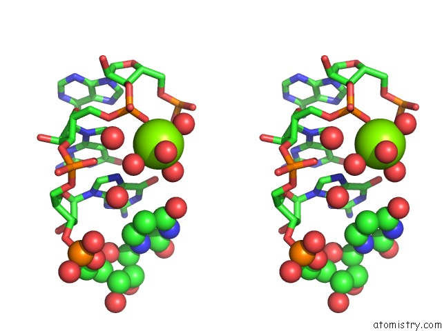
Magnesium binding site 1 out of 4 in 1dul
Go back to
Magnesium binding site 1 out
of 4 in the Structure of the Ribonucleoprotein Core of the E. Coli Signal Recognition Particle

Mono view
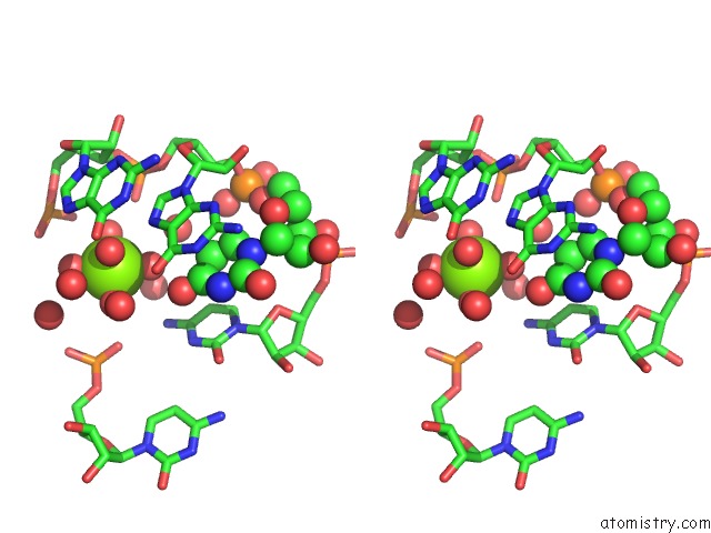
Stereo pair view
Mono view
Stereo pair view
A full contact list of Magnesium with other atoms in the Mg binding
site number 1 of Structure of the Ribonucleoprotein Core of the E. Coli Signal Recognition Particle within 5.0Å range:
|
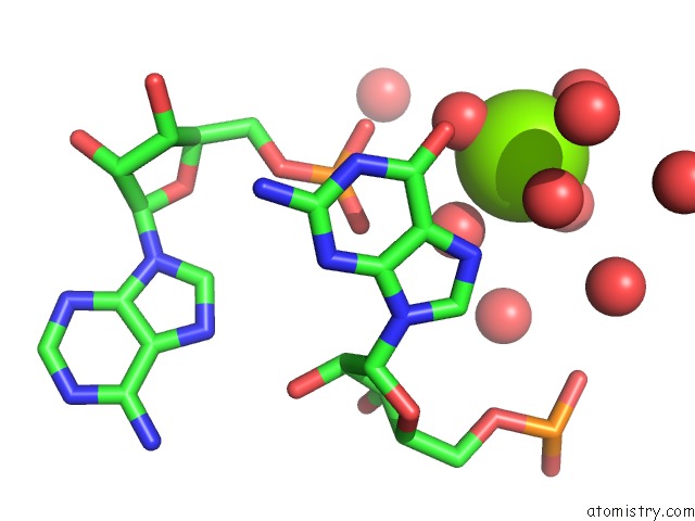
Magnesium binding site 2 out of 4 in 1dul
Go back to
Magnesium binding site 2 out
of 4 in the Structure of the Ribonucleoprotein Core of the E. Coli Signal Recognition Particle
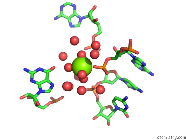
Mono view
Stereo pair view
Mono view
Stereo pair view
A full contact list of Magnesium with other atoms in the Mg binding
site number 2 of Structure of the Ribonucleoprotein Core of the E. Coli Signal Recognition Particle within 5.0Å range:
|
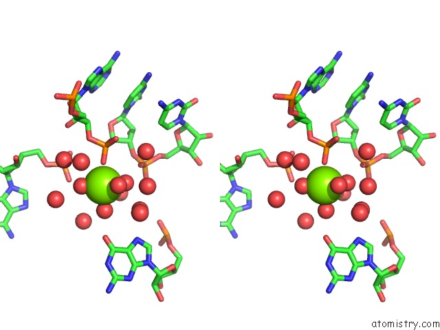
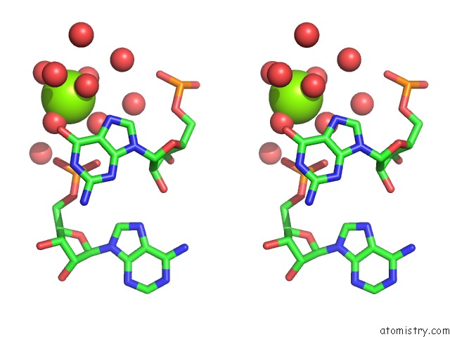
Magnesium binding site 3 out of 4 in 1dul
Go back to
Magnesium binding site 3 out
of 4 in the Structure of the Ribonucleoprotein Core of the E. Coli Signal Recognition Particle
Mono view
Stereo pair view
Mono view
Stereo pair view
A full contact list of Magnesium with other atoms in the Mg binding
site number 3 of Structure of the Ribonucleoprotein Core of the E. Coli Signal Recognition Particle within 5.0Å range:
|
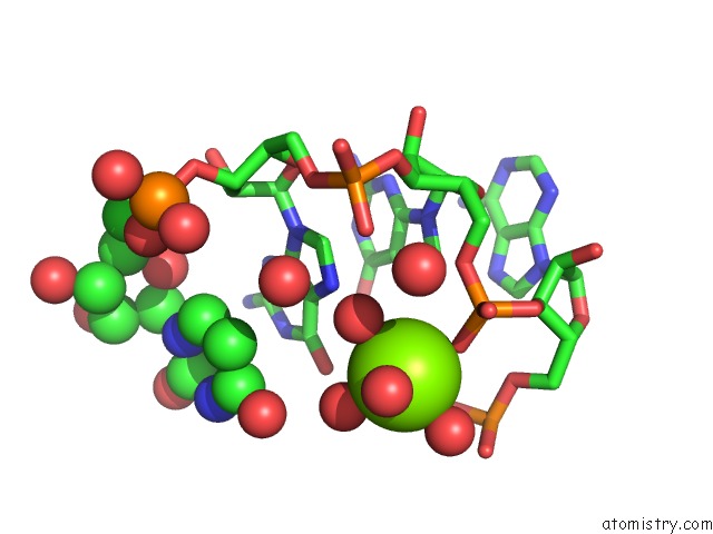
Magnesium binding site 4 out of 4 in 1dul
Go back to
Magnesium binding site 4 out
of 4 in the Structure of the Ribonucleoprotein Core of the E. Coli Signal Recognition Particle
Mono view
Stereo pair view
Mono view
Stereo pair view
A full contact list of Magnesium with other atoms in the Mg binding
site number 4 of Structure of the Ribonucleoprotein Core of the E. Coli Signal Recognition Particle within 5.0Å range:
|
Reference:
R.T.Batey,
R.P.Rambo,
L.Lucast,
B.Rha,
J.A.Doudna.
Crystal Structure of the Ribonucleoprotein Core of the Signal Recognition Particle. Science V. 287 1232 2000.
ISSN: ISSN 0036-8075
PubMed: 10678824
DOI: 10.1126/SCIENCE.287.5456.1232
Page generated: Tue Aug 13 02:45:51 2024
ISSN: ISSN 0036-8075
PubMed: 10678824
DOI: 10.1126/SCIENCE.287.5456.1232
Last articles
Fe in 2YXOFe in 2YRS
Fe in 2YXC
Fe in 2YNM
Fe in 2YVJ
Fe in 2YP1
Fe in 2YU2
Fe in 2YU1
Fe in 2YQB
Fe in 2YOO