Magnesium »
PDB 1duh-1e9a »
1e96 »
Magnesium in PDB 1e96: Structure of the Rac/P67PHOX Complex
Protein crystallography data
The structure of Structure of the Rac/P67PHOX Complex, PDB code: 1e96
was solved by
K.Lapouge,
S.J.M.Smith,
P.A.Walker,
S.J.Gamblin,
S.J.Smerdon,
K.Rittinger,
with X-Ray Crystallography technique. A brief refinement statistics is given in the table below:
| Resolution Low / High (Å) | 15.00 / 2.40 |
| Space group | P 32 2 1 |
| Cell size a, b, c (Å), α, β, γ (°) | 83.220, 83.220, 138.530, 90.00, 90.00, 120.00 |
| R / Rfree (%) | 24.4 / 28.4 |
Magnesium Binding Sites:
The binding sites of Magnesium atom in the Structure of the Rac/P67PHOX Complex
(pdb code 1e96). This binding sites where shown within
5.0 Angstroms radius around Magnesium atom.
In total only one binding site of Magnesium was determined in the Structure of the Rac/P67PHOX Complex, PDB code: 1e96:
In total only one binding site of Magnesium was determined in the Structure of the Rac/P67PHOX Complex, PDB code: 1e96:
Magnesium binding site 1 out of 1 in 1e96
Go back to
Magnesium binding site 1 out
of 1 in the Structure of the Rac/P67PHOX Complex
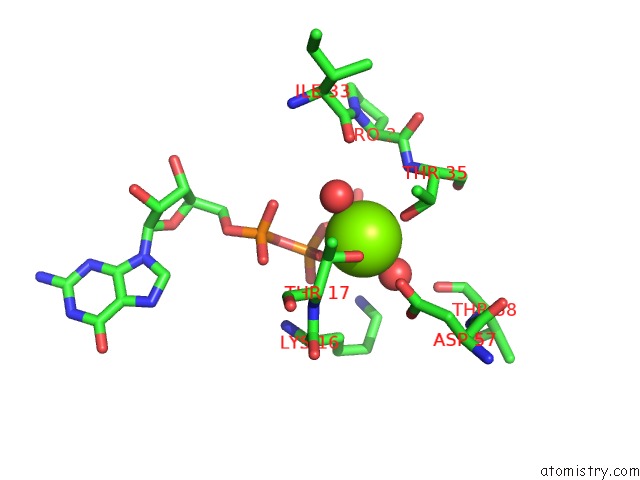
Mono view
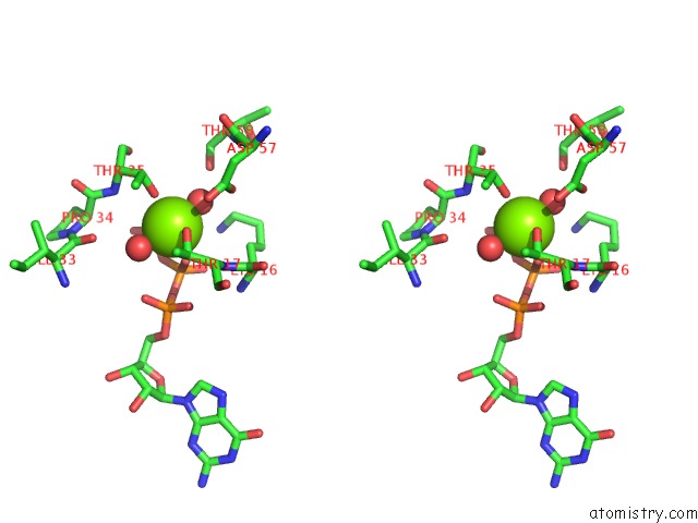
Stereo pair view
Mono view
Stereo pair view
A full contact list of Magnesium with other atoms in the Mg binding
site number 1 of Structure of the Rac/P67PHOX Complex within 5.0Å range:
|
Reference:
K.Lapouge,
S.J.Smith,
P.A.Walker,
S.J.Gamblin,
S.J.Smerdon,
K.Rittinger.
Structure of the Tpr Domain of P67PHOX in Complex with Rac.Gtp. Mol.Cell V. 6 899 2000.
ISSN: ISSN 1097-2765
PubMed: 11090627
DOI: 10.1016/S1097-2765(05)00091-2
Page generated: Tue Aug 13 02:52:09 2024
ISSN: ISSN 1097-2765
PubMed: 11090627
DOI: 10.1016/S1097-2765(05)00091-2
Last articles
Zn in 9J0NZn in 9J0O
Zn in 9J0P
Zn in 9FJX
Zn in 9EKB
Zn in 9C0F
Zn in 9CAH
Zn in 9CH0
Zn in 9CH3
Zn in 9CH1