Magnesium »
PDB 1e9b-1enn »
1ebg »
Magnesium in PDB 1ebg: Chelation of Ser 39 to MG2+ Latches A Gate at the Active Site of Enolase: Structure of the Bis(MG2+) Complex of Yeast Enolase and the Intermediate Analog Phosphonoacetohydroxamate at 2.1 Angstroms Resolution
Enzymatic activity of Chelation of Ser 39 to MG2+ Latches A Gate at the Active Site of Enolase: Structure of the Bis(MG2+) Complex of Yeast Enolase and the Intermediate Analog Phosphonoacetohydroxamate at 2.1 Angstroms Resolution
All present enzymatic activity of Chelation of Ser 39 to MG2+ Latches A Gate at the Active Site of Enolase: Structure of the Bis(MG2+) Complex of Yeast Enolase and the Intermediate Analog Phosphonoacetohydroxamate at 2.1 Angstroms Resolution:
4.2.1.11;
4.2.1.11;
Protein crystallography data
The structure of Chelation of Ser 39 to MG2+ Latches A Gate at the Active Site of Enolase: Structure of the Bis(MG2+) Complex of Yeast Enolase and the Intermediate Analog Phosphonoacetohydroxamate at 2.1 Angstroms Resolution, PDB code: 1ebg
was solved by
J.E.Wedekind,
G.H.Reed,
I.Rayment,
with X-Ray Crystallography technique. A brief refinement statistics is given in the table below:
| Resolution Low / High (Å) | N/A / 2.10 |
| Space group | C 1 2 1 |
| Cell size a, b, c (Å), α, β, γ (°) | 123.500, 73.900, 94.800, 90.00, 93.30, 90.00 |
| R / Rfree (%) | n/a / n/a |
Magnesium Binding Sites:
The binding sites of Magnesium atom in the Chelation of Ser 39 to MG2+ Latches A Gate at the Active Site of Enolase: Structure of the Bis(MG2+) Complex of Yeast Enolase and the Intermediate Analog Phosphonoacetohydroxamate at 2.1 Angstroms Resolution
(pdb code 1ebg). This binding sites where shown within
5.0 Angstroms radius around Magnesium atom.
In total 4 binding sites of Magnesium where determined in the Chelation of Ser 39 to MG2+ Latches A Gate at the Active Site of Enolase: Structure of the Bis(MG2+) Complex of Yeast Enolase and the Intermediate Analog Phosphonoacetohydroxamate at 2.1 Angstroms Resolution, PDB code: 1ebg:
Jump to Magnesium binding site number: 1; 2; 3; 4;
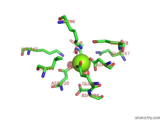
Magnesium binding site 1 out of 4 in 1ebg
Go back to
Magnesium binding site 1 out
of 4 in the Chelation of Ser 39 to MG2+ Latches A Gate at the Active Site of Enolase: Structure of the Bis(MG2+) Complex of Yeast Enolase and the Intermediate Analog Phosphonoacetohydroxamate at 2.1 Angstroms Resolution
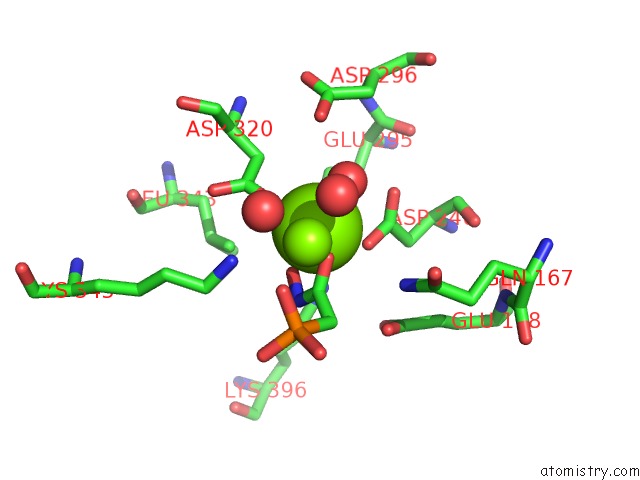
Mono view
Stereo pair view
Mono view
Stereo pair view
A full contact list of Magnesium with other atoms in the Mg binding
site number 1 of Chelation of Ser 39 to MG2+ Latches A Gate at the Active Site of Enolase: Structure of the Bis(MG2+) Complex of Yeast Enolase and the Intermediate Analog Phosphonoacetohydroxamate at 2.1 Angstroms Resolution within 5.0Å range:
|
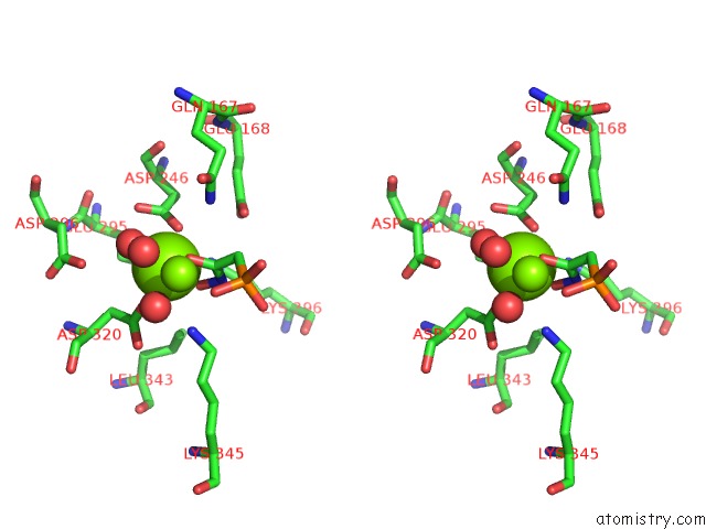
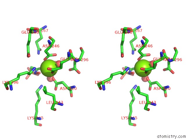
Magnesium binding site 2 out of 4 in 1ebg
Go back to
Magnesium binding site 2 out
of 4 in the Chelation of Ser 39 to MG2+ Latches A Gate at the Active Site of Enolase: Structure of the Bis(MG2+) Complex of Yeast Enolase and the Intermediate Analog Phosphonoacetohydroxamate at 2.1 Angstroms Resolution
Mono view
Stereo pair view
Mono view
Stereo pair view
A full contact list of Magnesium with other atoms in the Mg binding
site number 2 of Chelation of Ser 39 to MG2+ Latches A Gate at the Active Site of Enolase: Structure of the Bis(MG2+) Complex of Yeast Enolase and the Intermediate Analog Phosphonoacetohydroxamate at 2.1 Angstroms Resolution within 5.0Å range:
|
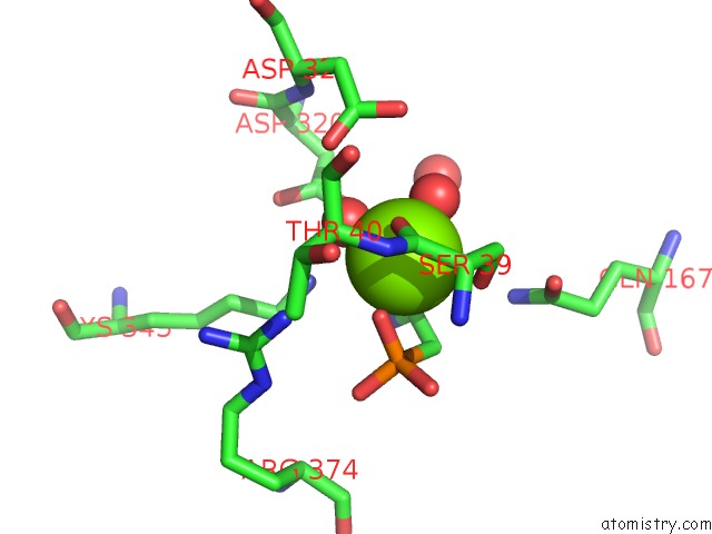
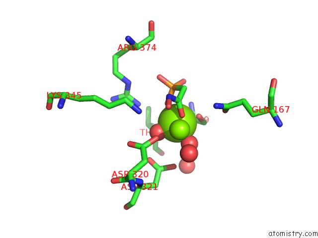
Magnesium binding site 3 out of 4 in 1ebg
Go back to
Magnesium binding site 3 out
of 4 in the Chelation of Ser 39 to MG2+ Latches A Gate at the Active Site of Enolase: Structure of the Bis(MG2+) Complex of Yeast Enolase and the Intermediate Analog Phosphonoacetohydroxamate at 2.1 Angstroms Resolution
Mono view
Stereo pair view
Mono view
Stereo pair view
A full contact list of Magnesium with other atoms in the Mg binding
site number 3 of Chelation of Ser 39 to MG2+ Latches A Gate at the Active Site of Enolase: Structure of the Bis(MG2+) Complex of Yeast Enolase and the Intermediate Analog Phosphonoacetohydroxamate at 2.1 Angstroms Resolution within 5.0Å range:
|
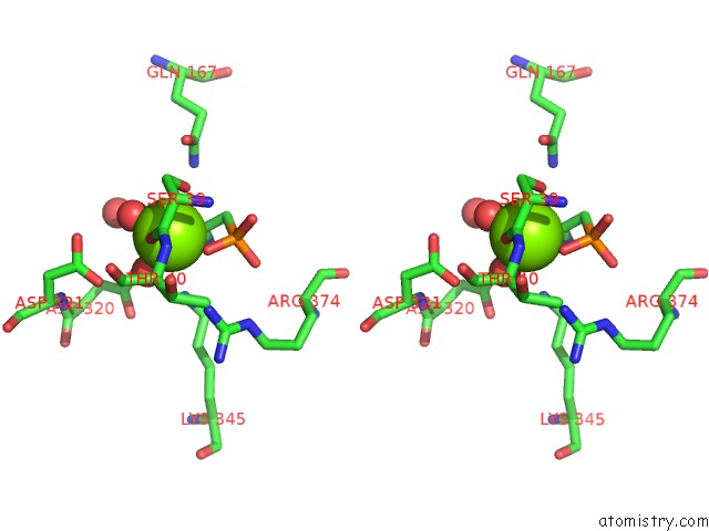
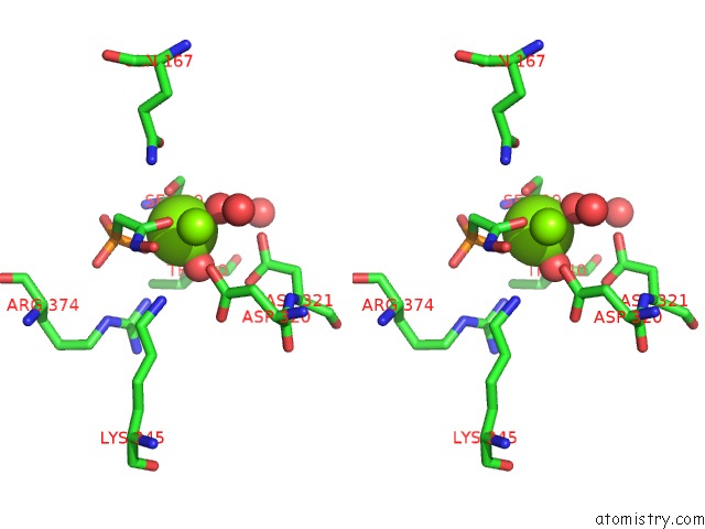
Magnesium binding site 4 out of 4 in 1ebg
Go back to
Magnesium binding site 4 out
of 4 in the Chelation of Ser 39 to MG2+ Latches A Gate at the Active Site of Enolase: Structure of the Bis(MG2+) Complex of Yeast Enolase and the Intermediate Analog Phosphonoacetohydroxamate at 2.1 Angstroms Resolution
Mono view
Stereo pair view
Mono view
Stereo pair view
A full contact list of Magnesium with other atoms in the Mg binding
site number 4 of Chelation of Ser 39 to MG2+ Latches A Gate at the Active Site of Enolase: Structure of the Bis(MG2+) Complex of Yeast Enolase and the Intermediate Analog Phosphonoacetohydroxamate at 2.1 Angstroms Resolution within 5.0Å range:
|
Reference:
J.E.Wedekind,
R.R.Poyner,
G.H.Reed,
I.Rayment.
Chelation of Serine 39 to MG2+ Latches A Gate at the Active Site of Enolase: Structure of the Bis(MG2+) Complex of Yeast Enolase and the Intermediate Analog Phosphonoacetohydroxamate at 2.1-A Resolution. Biochemistry V. 33 9333 1994.
ISSN: ISSN 0006-2960
PubMed: 8049235
DOI: 10.1021/BI00197A038
Page generated: Tue Aug 13 02:54:08 2024
ISSN: ISSN 0006-2960
PubMed: 8049235
DOI: 10.1021/BI00197A038
Last articles
Zn in 9MJ5Zn in 9HNW
Zn in 9G0L
Zn in 9FNE
Zn in 9DZN
Zn in 9E0I
Zn in 9D32
Zn in 9DAK
Zn in 8ZXC
Zn in 8ZUF