Magnesium »
PDB 1e9b-1enn »
1ec9 »
Magnesium in PDB 1ec9: E. Coli Glucarate Dehydratase Bound to Xylarohydroxamate
Enzymatic activity of E. Coli Glucarate Dehydratase Bound to Xylarohydroxamate
All present enzymatic activity of E. Coli Glucarate Dehydratase Bound to Xylarohydroxamate:
4.2.1.40;
4.2.1.40;
Protein crystallography data
The structure of E. Coli Glucarate Dehydratase Bound to Xylarohydroxamate, PDB code: 1ec9
was solved by
A.M.Gulick,
B.K.Hubbard,
J.A.Gerlt,
I.Rayment,
with X-Ray Crystallography technique. A brief refinement statistics is given in the table below:
| Resolution Low / High (Å) | 30.00 / 2.00 |
| Space group | P 1 |
| Cell size a, b, c (Å), α, β, γ (°) | 71.294, 84.836, 98.987, 103.14, 94.31, 113.20 |
| R / Rfree (%) | n/a / n/a |
Magnesium Binding Sites:
The binding sites of Magnesium atom in the E. Coli Glucarate Dehydratase Bound to Xylarohydroxamate
(pdb code 1ec9). This binding sites where shown within
5.0 Angstroms radius around Magnesium atom.
In total 4 binding sites of Magnesium where determined in the E. Coli Glucarate Dehydratase Bound to Xylarohydroxamate, PDB code: 1ec9:
Jump to Magnesium binding site number: 1; 2; 3; 4;
In total 4 binding sites of Magnesium where determined in the E. Coli Glucarate Dehydratase Bound to Xylarohydroxamate, PDB code: 1ec9:
Jump to Magnesium binding site number: 1; 2; 3; 4;
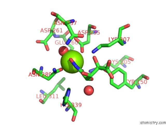
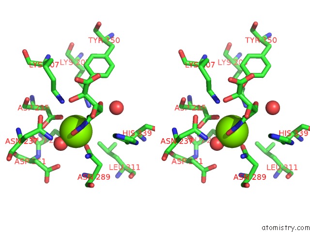
Magnesium binding site 1 out of 4 in 1ec9
Go back to
Magnesium binding site 1 out
of 4 in the E. Coli Glucarate Dehydratase Bound to Xylarohydroxamate
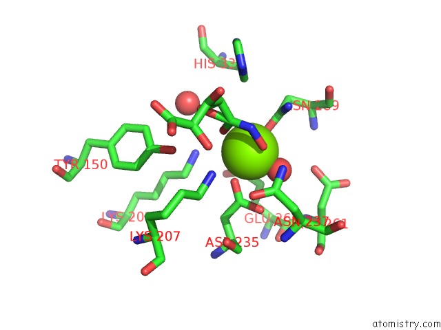
Mono view
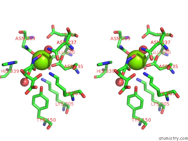
Stereo pair view
Mono view
Stereo pair view
A full contact list of Magnesium with other atoms in the Mg binding
site number 1 of E. Coli Glucarate Dehydratase Bound to Xylarohydroxamate within 5.0Å range:
|
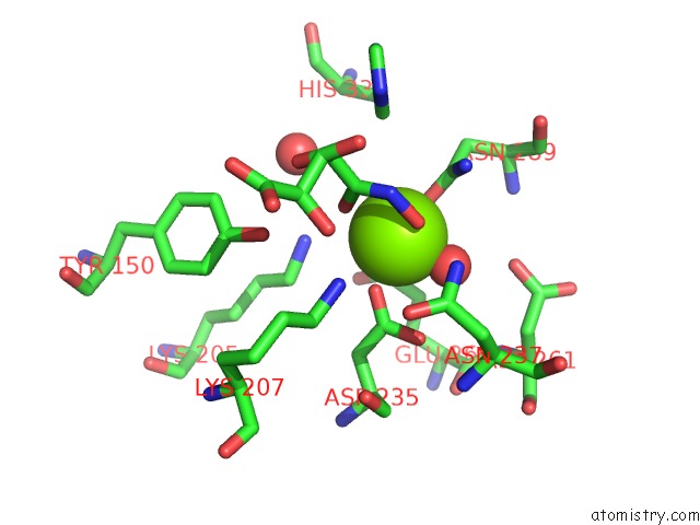
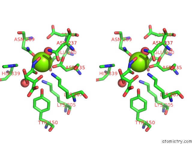
Magnesium binding site 2 out of 4 in 1ec9
Go back to
Magnesium binding site 2 out
of 4 in the E. Coli Glucarate Dehydratase Bound to Xylarohydroxamate
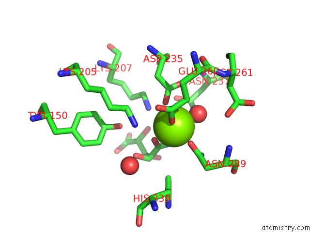
Mono view
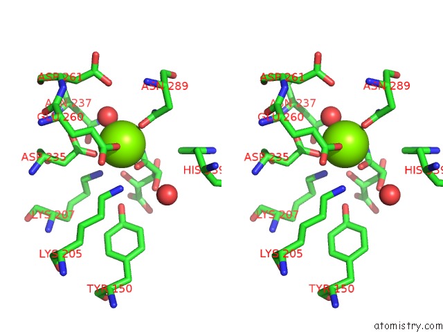
Stereo pair view
Mono view
Stereo pair view
A full contact list of Magnesium with other atoms in the Mg binding
site number 2 of E. Coli Glucarate Dehydratase Bound to Xylarohydroxamate within 5.0Å range:
|
Magnesium binding site 3 out of 4 in 1ec9
Go back to
Magnesium binding site 3 out
of 4 in the E. Coli Glucarate Dehydratase Bound to Xylarohydroxamate
Mono view
Stereo pair view
Mono view
Stereo pair view
A full contact list of Magnesium with other atoms in the Mg binding
site number 3 of E. Coli Glucarate Dehydratase Bound to Xylarohydroxamate within 5.0Å range:
|
Magnesium binding site 4 out of 4 in 1ec9
Go back to
Magnesium binding site 4 out
of 4 in the E. Coli Glucarate Dehydratase Bound to Xylarohydroxamate
Mono view
Stereo pair view
Mono view
Stereo pair view
A full contact list of Magnesium with other atoms in the Mg binding
site number 4 of E. Coli Glucarate Dehydratase Bound to Xylarohydroxamate within 5.0Å range:
|
Reference:
A.M.Gulick,
B.K.Hubbard,
J.A.Gerlt,
I.Rayment.
Evolution of Enzymatic Activities in the Enolase Superfamily: Crystallographic and Mutagenesis Studies of the Reaction Catalyzed By D-Glucarate Dehydratase From Escherichia Coli. Biochemistry V. 39 4590 2000.
ISSN: ISSN 0006-2960
PubMed: 10769114
DOI: 10.1021/BI992782I
Page generated: Tue Aug 13 02:54:53 2024
ISSN: ISSN 0006-2960
PubMed: 10769114
DOI: 10.1021/BI992782I
Last articles
Fe in 2YXOFe in 2YRS
Fe in 2YXC
Fe in 2YNM
Fe in 2YVJ
Fe in 2YP1
Fe in 2YU2
Fe in 2YU1
Fe in 2YQB
Fe in 2YOO