Magnesium »
PDB 1e9b-1enn »
1eeo »
Magnesium in PDB 1eeo: Crystal Structure of Protein Tyrosine Phosphatase 1B Complexed with Acetyl-E-L-E-F-Ptyr-M-D-Y-E-NH2
Enzymatic activity of Crystal Structure of Protein Tyrosine Phosphatase 1B Complexed with Acetyl-E-L-E-F-Ptyr-M-D-Y-E-NH2
All present enzymatic activity of Crystal Structure of Protein Tyrosine Phosphatase 1B Complexed with Acetyl-E-L-E-F-Ptyr-M-D-Y-E-NH2:
3.1.3.48;
3.1.3.48;
Protein crystallography data
The structure of Crystal Structure of Protein Tyrosine Phosphatase 1B Complexed with Acetyl-E-L-E-F-Ptyr-M-D-Y-E-NH2, PDB code: 1eeo
was solved by
M.Sarmiento,
Y.A.Puius,
S.W.Vetter,
D.S.Lawrence,
S.C.Almo,
Z.Y.Zhang,
with X-Ray Crystallography technique. A brief refinement statistics is given in the table below:
| Resolution Low / High (Å) | 24.09 / 1.80 |
| Space group | P 21 21 21 |
| Cell size a, b, c (Å), α, β, γ (°) | 52.581, 82.050, 88.543, 90.00, 90.00, 90.00 |
| R / Rfree (%) | 18.3 / 21.2 |
Magnesium Binding Sites:
The binding sites of Magnesium atom in the Crystal Structure of Protein Tyrosine Phosphatase 1B Complexed with Acetyl-E-L-E-F-Ptyr-M-D-Y-E-NH2
(pdb code 1eeo). This binding sites where shown within
5.0 Angstroms radius around Magnesium atom.
In total only one binding site of Magnesium was determined in the Crystal Structure of Protein Tyrosine Phosphatase 1B Complexed with Acetyl-E-L-E-F-Ptyr-M-D-Y-E-NH2, PDB code: 1eeo:
In total only one binding site of Magnesium was determined in the Crystal Structure of Protein Tyrosine Phosphatase 1B Complexed with Acetyl-E-L-E-F-Ptyr-M-D-Y-E-NH2, PDB code: 1eeo:
Magnesium binding site 1 out of 1 in 1eeo
Go back to
Magnesium binding site 1 out
of 1 in the Crystal Structure of Protein Tyrosine Phosphatase 1B Complexed with Acetyl-E-L-E-F-Ptyr-M-D-Y-E-NH2
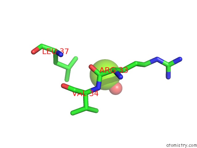
Mono view
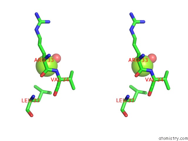
Stereo pair view
Mono view
Stereo pair view
A full contact list of Magnesium with other atoms in the Mg binding
site number 1 of Crystal Structure of Protein Tyrosine Phosphatase 1B Complexed with Acetyl-E-L-E-F-Ptyr-M-D-Y-E-NH2 within 5.0Å range:
|
Reference:
M.Sarmiento,
Y.A.Puius,
S.W.Vetter,
Y.F.Keng,
L.Wu,
Y.Zhao,
D.S.Lawrence,
S.C.Almo,
Z.Y.Zhang.
Structural Basis of Plasticity in Protein Tyrosine Phosphatase 1B Substrate Recognition. Biochemistry V. 39 8171 2000.
ISSN: ISSN 0006-2960
PubMed: 10889023
DOI: 10.1021/BI000319W
Page generated: Tue Aug 13 02:56:56 2024
ISSN: ISSN 0006-2960
PubMed: 10889023
DOI: 10.1021/BI000319W
Last articles
Fe in 2YXOFe in 2YRS
Fe in 2YXC
Fe in 2YNM
Fe in 2YVJ
Fe in 2YP1
Fe in 2YU2
Fe in 2YU1
Fe in 2YQB
Fe in 2YOO