Magnesium »
PDB 1eo3-1f6t »
1esq »
Magnesium in PDB 1esq: Crystal Structure of Thiazole Kinase Mutant (C198S) with Atp and Thiazole Phosphate.
Enzymatic activity of Crystal Structure of Thiazole Kinase Mutant (C198S) with Atp and Thiazole Phosphate.
All present enzymatic activity of Crystal Structure of Thiazole Kinase Mutant (C198S) with Atp and Thiazole Phosphate.:
2.7.1.50;
2.7.1.50;
Protein crystallography data
The structure of Crystal Structure of Thiazole Kinase Mutant (C198S) with Atp and Thiazole Phosphate., PDB code: 1esq
was solved by
N.Campobasso,
I.I.Mathews,
T.P.Begley,
S.E.Ealick,
with X-Ray Crystallography technique. A brief refinement statistics is given in the table below:
| Resolution Low / High (Å) | 20.00 / 2.50 |
| Space group | P 1 21 1 |
| Cell size a, b, c (Å), α, β, γ (°) | 54.080, 100.840, 72.510, 90.00, 95.10, 90.00 |
| R / Rfree (%) | 20.9 / 28.2 |
Magnesium Binding Sites:
The binding sites of Magnesium atom in the Crystal Structure of Thiazole Kinase Mutant (C198S) with Atp and Thiazole Phosphate.
(pdb code 1esq). This binding sites where shown within
5.0 Angstroms radius around Magnesium atom.
In total 6 binding sites of Magnesium where determined in the Crystal Structure of Thiazole Kinase Mutant (C198S) with Atp and Thiazole Phosphate., PDB code: 1esq:
Jump to Magnesium binding site number: 1; 2; 3; 4; 5; 6;
In total 6 binding sites of Magnesium where determined in the Crystal Structure of Thiazole Kinase Mutant (C198S) with Atp and Thiazole Phosphate., PDB code: 1esq:
Jump to Magnesium binding site number: 1; 2; 3; 4; 5; 6;
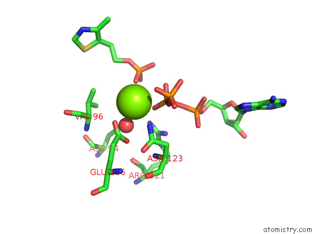
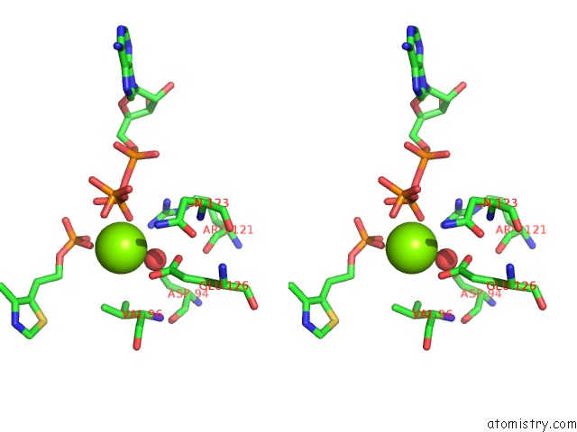
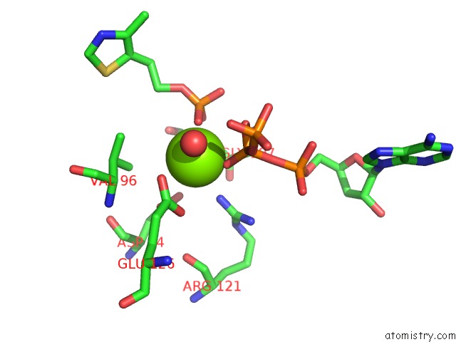
Magnesium binding site 1 out of 6 in 1esq
Go back to
Magnesium binding site 1 out
of 6 in the Crystal Structure of Thiazole Kinase Mutant (C198S) with Atp and Thiazole Phosphate.
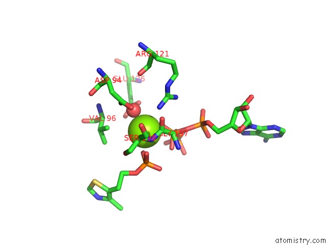
Mono view
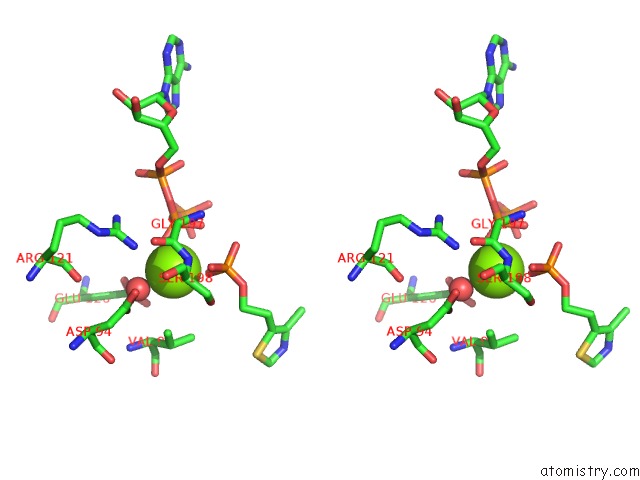
Stereo pair view
Mono view
Stereo pair view
A full contact list of Magnesium with other atoms in the Mg binding
site number 1 of Crystal Structure of Thiazole Kinase Mutant (C198S) with Atp and Thiazole Phosphate. within 5.0Å range:
|
Magnesium binding site 2 out of 6 in 1esq
Go back to
Magnesium binding site 2 out
of 6 in the Crystal Structure of Thiazole Kinase Mutant (C198S) with Atp and Thiazole Phosphate.
Mono view
Stereo pair view
Mono view
Stereo pair view
A full contact list of Magnesium with other atoms in the Mg binding
site number 2 of Crystal Structure of Thiazole Kinase Mutant (C198S) with Atp and Thiazole Phosphate. within 5.0Å range:
|
Magnesium binding site 3 out of 6 in 1esq
Go back to
Magnesium binding site 3 out
of 6 in the Crystal Structure of Thiazole Kinase Mutant (C198S) with Atp and Thiazole Phosphate.
Mono view
Stereo pair view
Mono view
Stereo pair view
A full contact list of Magnesium with other atoms in the Mg binding
site number 3 of Crystal Structure of Thiazole Kinase Mutant (C198S) with Atp and Thiazole Phosphate. within 5.0Å range:
|
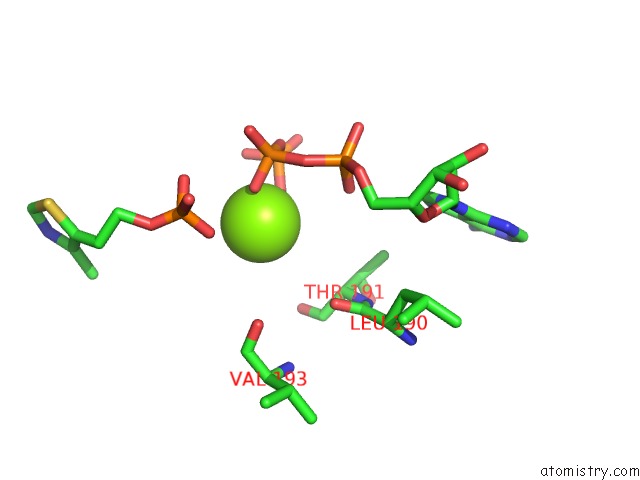
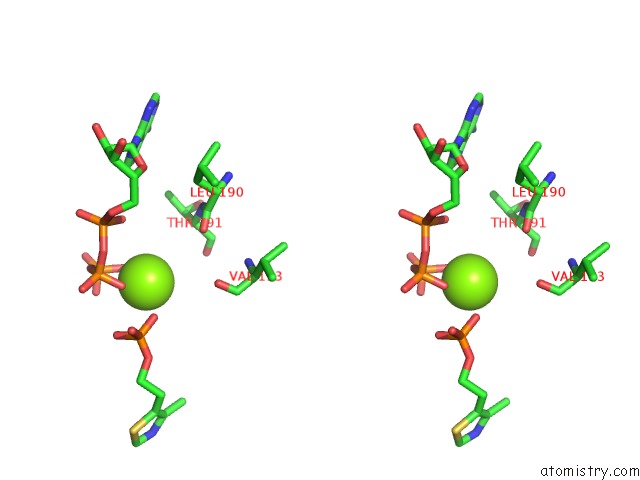
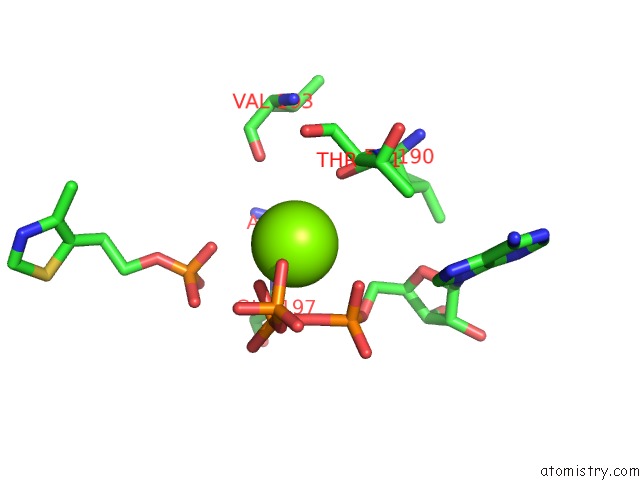
Magnesium binding site 4 out of 6 in 1esq
Go back to
Magnesium binding site 4 out
of 6 in the Crystal Structure of Thiazole Kinase Mutant (C198S) with Atp and Thiazole Phosphate.
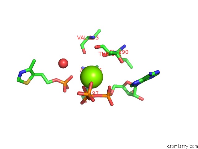
Mono view
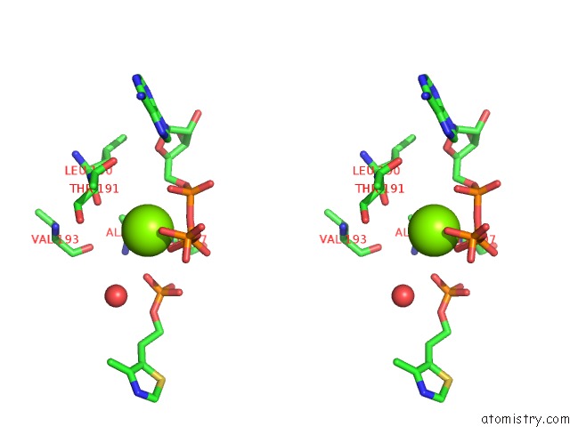
Stereo pair view
Mono view
Stereo pair view
A full contact list of Magnesium with other atoms in the Mg binding
site number 4 of Crystal Structure of Thiazole Kinase Mutant (C198S) with Atp and Thiazole Phosphate. within 5.0Å range:
|
Magnesium binding site 5 out of 6 in 1esq
Go back to
Magnesium binding site 5 out
of 6 in the Crystal Structure of Thiazole Kinase Mutant (C198S) with Atp and Thiazole Phosphate.
Mono view

Stereo pair view
Mono view
Stereo pair view
A full contact list of Magnesium with other atoms in the Mg binding
site number 5 of Crystal Structure of Thiazole Kinase Mutant (C198S) with Atp and Thiazole Phosphate. within 5.0Å range:
|
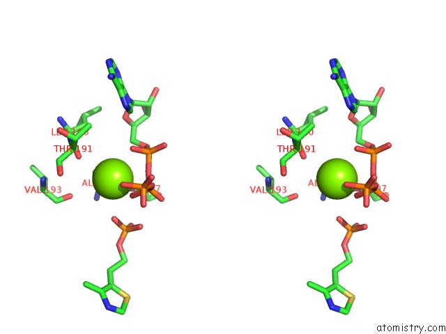
Magnesium binding site 6 out of 6 in 1esq
Go back to
Magnesium binding site 6 out
of 6 in the Crystal Structure of Thiazole Kinase Mutant (C198S) with Atp and Thiazole Phosphate.
Mono view
Stereo pair view
Mono view
Stereo pair view
A full contact list of Magnesium with other atoms in the Mg binding
site number 6 of Crystal Structure of Thiazole Kinase Mutant (C198S) with Atp and Thiazole Phosphate. within 5.0Å range:
|
Reference:
N.Campobasso,
I.I.Mathews,
T.P.Begley,
S.E.Ealick.
Crystal Structure of 4-Methyl-5-Beta-Hydroxyethylthiazole Kinase From Bacillus Subtilis at 1.5 A Resolution. Biochemistry V. 39 7868 2000.
ISSN: ISSN 0006-2960
PubMed: 10891066
DOI: 10.1021/BI0000061
Page generated: Sat Aug 9 20:48:04 2025
ISSN: ISSN 0006-2960
PubMed: 10891066
DOI: 10.1021/BI0000061
Last articles
Mg in 3CR1Mg in 3CME
Mg in 3CMA
Mg in 3CP6
Mg in 3CPJ
Mg in 3CPH
Mg in 3CON
Mg in 3CMV
Mg in 3COB
Mg in 3CNZ