Magnesium in PDB 1eyz: Structure of Escherichia Coli Purt-Encoded Glycinamide Ribonucleotide Transformylase Complexed with Mg and Amppnp
Protein crystallography data
The structure of Structure of Escherichia Coli Purt-Encoded Glycinamide Ribonucleotide Transformylase Complexed with Mg and Amppnp, PDB code: 1eyz
was solved by
J.B.Thoden,
S.Firestine,
A.Nixon,
S.J.Benkovic,
H.M.Holden,
with X-Ray Crystallography technique. A brief refinement statistics is given in the table below:
Resolution Low / High (Å)
|
30.00 /
1.75
|
Space group
|
P 21 21 2
|
Cell size a, b, c (Å), α, β, γ (°)
|
62.300,
179.500,
75.700,
90.00,
90.00,
90.00
|
R / Rfree (%)
|
n/a /
n/a
|
Other elements in 1eyz:
The structure of Structure of Escherichia Coli Purt-Encoded Glycinamide Ribonucleotide Transformylase Complexed with Mg and Amppnp also contains other interesting chemical elements:
Magnesium Binding Sites:
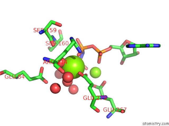
The binding sites of Magnesium atom in the Structure of Escherichia Coli Purt-Encoded Glycinamide Ribonucleotide Transformylase Complexed with Mg and Amppnp
(pdb code 1eyz). This binding sites where shown within
5.0 Angstroms radius around Magnesium atom.
In total 5 binding sites of Magnesium where determined in the
Structure of Escherichia Coli Purt-Encoded Glycinamide Ribonucleotide Transformylase Complexed with Mg and Amppnp, PDB code: 1eyz:
Jump to Magnesium binding site number:
1;
2;
3;
4;
5;
Magnesium binding site 1 out
of 5 in 1eyz
Go back to
Magnesium Binding Sites List in 1eyz
Magnesium binding site 1 out
of 5 in the Structure of Escherichia Coli Purt-Encoded Glycinamide Ribonucleotide Transformylase Complexed with Mg and Amppnp
Mono view
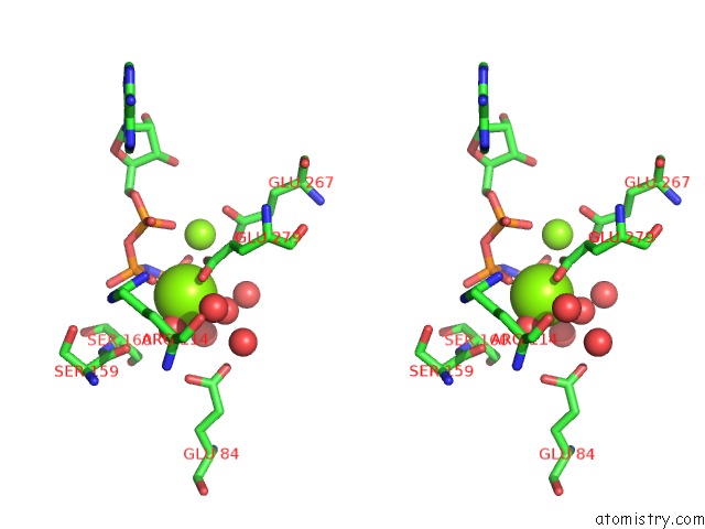
Stereo pair view
|
A full contact list of Magnesium with other atoms in the Mg binding
site number 1 of Structure of Escherichia Coli Purt-Encoded Glycinamide Ribonucleotide Transformylase Complexed with Mg and Amppnp within 5.0Å range:
probe
|
atom
|
residue
|
distance (Å)
|
B
|
Occ
|
A:Mg401
b:18.5
occ:1.00
|
O3G
|
A:ANP400
|
1.9
|
16.7
|
1.0
|
O2B
|
A:ANP400
|
2.0
|
15.8
|
1.0
|
O
|
A:HOH1119
|
2.1
|
15.6
|
1.0
|
OE1
|
A:GLU279
|
2.2
|
15.6
|
1.0
|
OE2
|
A:GLU279
|
2.3
|
16.1
|
1.0
|
O
|
A:HOH1131
|
2.3
|
23.1
|
1.0
|
CD
|
A:GLU279
|
2.5
|
15.3
|
1.0
|
PG
|
A:ANP400
|
3.2
|
18.0
|
1.0
|
PB
|
A:ANP400
|
3.2
|
16.7
|
1.0
|
N3B
|
A:ANP400
|
3.5
|
22.2
|
1.0
|
O2G
|
A:ANP400
|
3.6
|
13.3
|
1.0
|
MG
|
A:MG402
|
3.8
|
15.4
|
1.0
|
O
|
A:HOH1229
|
3.8
|
20.0
|
1.0
|
O2A
|
A:ANP400
|
3.9
|
15.8
|
1.0
|
O
|
A:HOH1175
|
4.0
|
22.5
|
1.0
|
CG
|
A:GLU279
|
4.0
|
9.4
|
1.0
|
NH1
|
A:ARG114
|
4.1
|
13.8
|
1.0
|
O
|
A:HOH1824
|
4.2
|
49.4
|
1.0
|
O1B
|
A:ANP400
|
4.2
|
21.6
|
1.0
|
O3A
|
A:ANP400
|
4.3
|
13.5
|
1.0
|
O
|
A:HOH1149
|
4.4
|
15.5
|
1.0
|
O
|
A:SER159
|
4.4
|
38.3
|
1.0
|
NH2
|
A:ARG114
|
4.4
|
16.9
|
1.0
|
O1G
|
A:ANP400
|
4.4
|
12.6
|
1.0
|
OE1
|
A:GLU84
|
4.5
|
28.7
|
1.0
|
PA
|
A:ANP400
|
4.5
|
13.9
|
1.0
|
OE2
|
A:GLU84
|
4.5
|
24.4
|
1.0
|
O1A
|
A:ANP400
|
4.6
|
13.6
|
1.0
|
CZ
|
A:ARG114
|
4.7
|
11.7
|
1.0
|
CA
|
A:SER160
|
4.7
|
30.1
|
1.0
|
OE1
|
A:GLU267
|
4.8
|
14.8
|
1.0
|
CB
|
A:SER160
|
4.8
|
87.5
|
1.0
|
CB
|
A:GLU279
|
4.9
|
8.6
|
1.0
|
CD
|
A:GLU84
|
5.0
|
0.0
|
1.0
|
|
Magnesium binding site 2 out
of 5 in 1eyz
Go back to
Magnesium Binding Sites List in 1eyz
Magnesium binding site 2 out
of 5 in the Structure of Escherichia Coli Purt-Encoded Glycinamide Ribonucleotide Transformylase Complexed with Mg and Amppnp
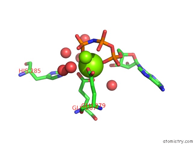
Mono view
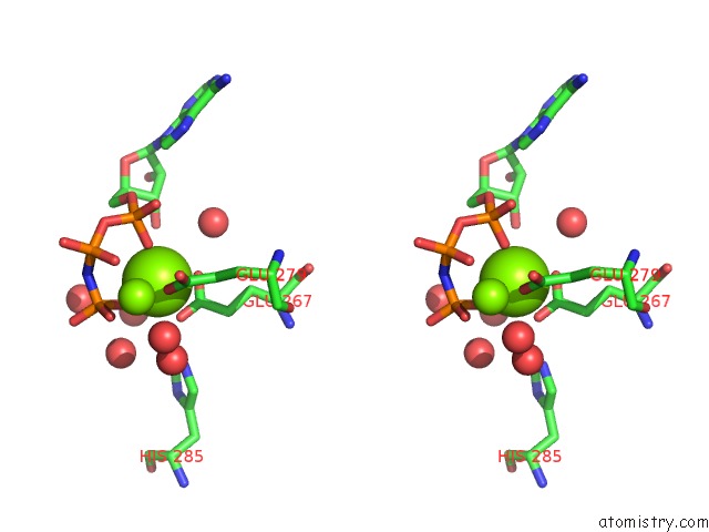
Stereo pair view
|
A full contact list of Magnesium with other atoms in the Mg binding
site number 2 of Structure of Escherichia Coli Purt-Encoded Glycinamide Ribonucleotide Transformylase Complexed with Mg and Amppnp within 5.0Å range:
probe
|
atom
|
residue
|
distance (Å)
|
B
|
Occ
|
A:Mg402
b:15.4
occ:1.00
|
O2A
|
A:ANP400
|
2.0
|
15.8
|
1.0
|
O2G
|
A:ANP400
|
2.0
|
13.3
|
1.0
|
OE1
|
A:GLU279
|
2.1
|
15.6
|
1.0
|
OE2
|
A:GLU267
|
2.1
|
14.1
|
1.0
|
O
|
A:HOH1113
|
2.2
|
15.1
|
1.0
|
OE1
|
A:GLU267
|
2.2
|
14.8
|
1.0
|
CD
|
A:GLU267
|
2.5
|
10.2
|
1.0
|
CD
|
A:GLU279
|
3.1
|
15.3
|
1.0
|
PG
|
A:ANP400
|
3.2
|
18.0
|
1.0
|
PA
|
A:ANP400
|
3.5
|
13.9
|
1.0
|
N3B
|
A:ANP400
|
3.6
|
22.2
|
1.0
|
CG
|
A:GLU279
|
3.7
|
9.4
|
1.0
|
O3G
|
A:ANP400
|
3.7
|
16.7
|
1.0
|
MG
|
A:MG401
|
3.8
|
18.5
|
1.0
|
CG
|
A:GLU267
|
4.0
|
13.1
|
1.0
|
O2B
|
A:ANP400
|
4.0
|
15.8
|
1.0
|
O
|
A:HOH1143
|
4.2
|
26.3
|
1.0
|
O3A
|
A:ANP400
|
4.2
|
13.5
|
1.0
|
OE2
|
A:GLU279
|
4.2
|
16.1
|
1.0
|
PB
|
A:ANP400
|
4.2
|
16.7
|
1.0
|
O
|
A:HOH1229
|
4.2
|
20.0
|
1.0
|
O1A
|
A:ANP400
|
4.2
|
13.6
|
1.0
|
O
|
A:HOH1331
|
4.3
|
41.2
|
1.0
|
O
|
A:HOH1174
|
4.3
|
15.5
|
1.0
|
O5'
|
A:ANP400
|
4.3
|
18.3
|
1.0
|
O3'
|
A:ANP400
|
4.4
|
13.6
|
1.0
|
O1G
|
A:ANP400
|
4.5
|
12.6
|
1.0
|
C5'
|
A:ANP400
|
4.6
|
19.4
|
1.0
|
O
|
A:HOH1131
|
4.6
|
23.1
|
1.0
|
O
|
A:HOH1258
|
4.6
|
32.7
|
1.0
|
NE2
|
A:HIS285
|
4.8
|
17.4
|
1.0
|
CB
|
A:GLU267
|
4.8
|
10.2
|
1.0
|
|
Magnesium binding site 3 out
of 5 in 1eyz
Go back to
Magnesium Binding Sites List in 1eyz
Magnesium binding site 3 out
of 5 in the Structure of Escherichia Coli Purt-Encoded Glycinamide Ribonucleotide Transformylase Complexed with Mg and Amppnp
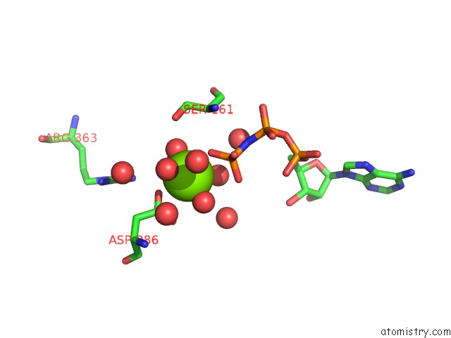
Mono view
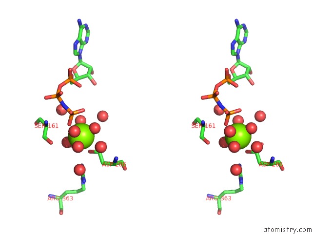
Stereo pair view
|
A full contact list of Magnesium with other atoms in the Mg binding
site number 3 of Structure of Escherichia Coli Purt-Encoded Glycinamide Ribonucleotide Transformylase Complexed with Mg and Amppnp within 5.0Å range:
probe
|
atom
|
residue
|
distance (Å)
|
B
|
Occ
|
A:Mg403
b:29.6
occ:1.00
|
O
|
A:HOH1143
|
2.1
|
26.3
|
1.0
|
O
|
A:HOH1792
|
2.2
|
29.5
|
1.0
|
O
|
A:HOH1794
|
2.3
|
30.6
|
1.0
|
O1G
|
A:ANP400
|
2.3
|
12.6
|
1.0
|
OD2
|
A:ASP286
|
2.6
|
31.0
|
1.0
|
O
|
A:HOH1258
|
3.0
|
32.7
|
1.0
|
CG
|
A:ASP286
|
3.2
|
14.2
|
1.0
|
OD1
|
A:ASP286
|
3.3
|
14.8
|
1.0
|
PG
|
A:ANP400
|
3.4
|
18.0
|
1.0
|
O2G
|
A:ANP400
|
3.5
|
13.3
|
1.0
|
O
|
A:HOH1394
|
3.6
|
38.0
|
1.0
|
O
|
A:HOH1144
|
3.8
|
19.3
|
1.0
|
OG
|
A:SER161
|
4.0
|
63.2
|
1.0
|
O
|
A:HOH1175
|
4.2
|
22.5
|
1.0
|
O
|
A:HOH1331
|
4.3
|
41.2
|
1.0
|
O3G
|
A:ANP400
|
4.4
|
16.7
|
1.0
|
O
|
A:HOH1173
|
4.5
|
23.6
|
1.0
|
CB
|
A:ASP286
|
4.5
|
15.2
|
1.0
|
N3B
|
A:ANP400
|
4.6
|
22.2
|
1.0
|
NH2
|
A:ARG363
|
4.6
|
23.8
|
1.0
|
CA
|
A:SER161
|
4.8
|
26.4
|
1.0
|
O
|
A:HOH1138
|
4.8
|
33.7
|
1.0
|
CB
|
A:SER161
|
4.9
|
71.4
|
1.0
|
N
|
A:SER161
|
4.9
|
27.6
|
1.0
|
|
Magnesium binding site 4 out
of 5 in 1eyz
Go back to
Magnesium Binding Sites List in 1eyz
Magnesium binding site 4 out
of 5 in the Structure of Escherichia Coli Purt-Encoded Glycinamide Ribonucleotide Transformylase Complexed with Mg and Amppnp
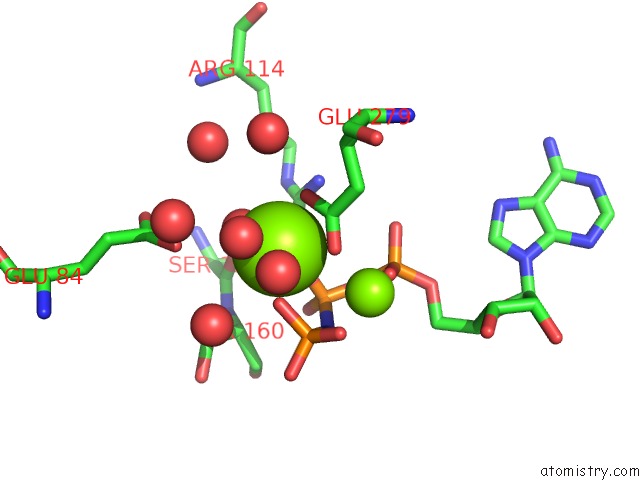
Mono view
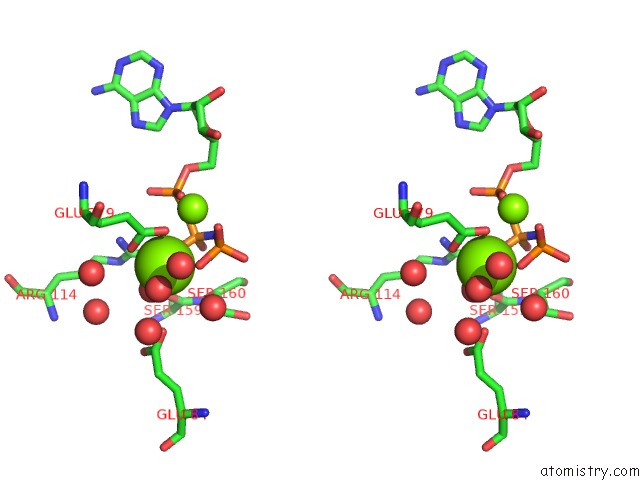
Stereo pair view
|
A full contact list of Magnesium with other atoms in the Mg binding
site number 4 of Structure of Escherichia Coli Purt-Encoded Glycinamide Ribonucleotide Transformylase Complexed with Mg and Amppnp within 5.0Å range:
probe
|
atom
|
residue
|
distance (Å)
|
B
|
Occ
|
B:Mg401
b:25.1
occ:1.00
|
O
|
B:HOH1615
|
1.9
|
31.6
|
1.0
|
O2B
|
B:ANP961
|
2.0
|
35.7
|
1.0
|
O3G
|
B:ANP961
|
2.0
|
24.7
|
1.0
|
OE2
|
B:GLU279
|
2.2
|
22.4
|
1.0
|
OE1
|
B:GLU279
|
2.4
|
28.1
|
1.0
|
O
|
B:HOH1885
|
2.5
|
32.5
|
1.0
|
CD
|
B:GLU279
|
2.6
|
25.1
|
1.0
|
PB
|
B:ANP961
|
3.3
|
37.8
|
1.0
|
PG
|
B:ANP961
|
3.3
|
36.6
|
1.0
|
N3B
|
B:ANP961
|
3.7
|
36.3
|
1.0
|
O
|
B:HOH1626
|
3.8
|
49.8
|
1.0
|
OE1
|
B:GLU84
|
3.8
|
47.9
|
1.0
|
O
|
B:HOH1542
|
3.8
|
27.1
|
1.0
|
NH2
|
B:ARG114
|
3.8
|
40.0
|
1.0
|
MG
|
B:MG402
|
3.9
|
23.9
|
1.0
|
O2G
|
B:ANP961
|
3.9
|
23.7
|
1.0
|
NH1
|
B:ARG114
|
4.1
|
25.6
|
1.0
|
O2A
|
B:ANP961
|
4.1
|
33.2
|
1.0
|
CG
|
B:GLU279
|
4.1
|
19.3
|
1.0
|
O3A
|
B:ANP961
|
4.2
|
18.6
|
1.0
|
O
|
B:HOH1661
|
4.2
|
34.6
|
1.0
|
O
|
B:SER159
|
4.3
|
87.0
|
1.0
|
O1B
|
B:ANP961
|
4.3
|
36.9
|
1.0
|
CZ
|
B:ARG114
|
4.3
|
26.9
|
1.0
|
O1G
|
B:ANP961
|
4.5
|
50.7
|
1.0
|
PA
|
B:ANP961
|
4.6
|
27.9
|
1.0
|
O
|
B:HOH1528
|
4.6
|
30.4
|
1.0
|
O1A
|
B:ANP961
|
4.7
|
20.2
|
1.0
|
CA
|
B:SER160
|
4.8
|
75.9
|
1.0
|
CD
|
B:GLU84
|
4.9
|
21.3
|
1.0
|
CB
|
B:SER160
|
4.9
|
41.5
|
1.0
|
O
|
B:HOH1505
|
4.9
|
24.4
|
1.0
|
CB
|
B:GLU279
|
4.9
|
13.3
|
1.0
|
|
Magnesium binding site 5 out
of 5 in 1eyz
Go back to
Magnesium Binding Sites List in 1eyz
Magnesium binding site 5 out
of 5 in the Structure of Escherichia Coli Purt-Encoded Glycinamide Ribonucleotide Transformylase Complexed with Mg and Amppnp
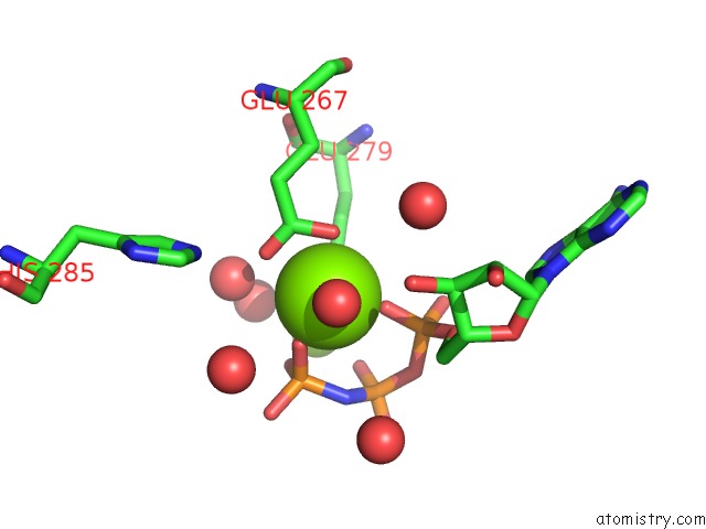
Mono view
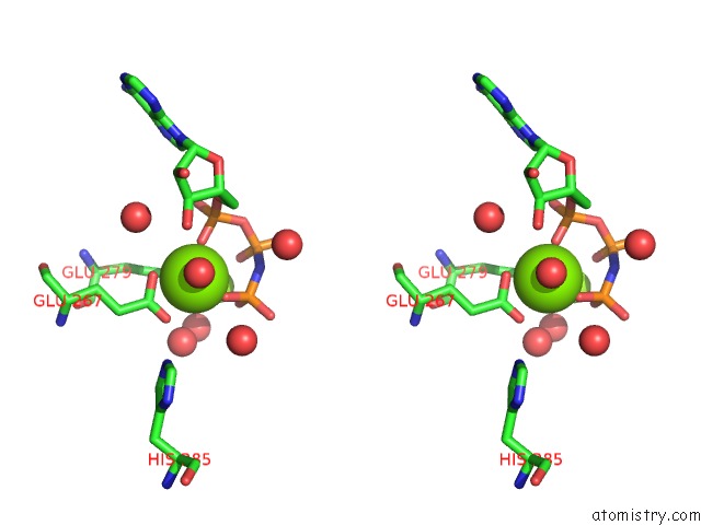
Stereo pair view
|
A full contact list of Magnesium with other atoms in the Mg binding
site number 5 of Structure of Escherichia Coli Purt-Encoded Glycinamide Ribonucleotide Transformylase Complexed with Mg and Amppnp within 5.0Å range:
probe
|
atom
|
residue
|
distance (Å)
|
B
|
Occ
|
B:Mg402
b:23.9
occ:1.00
|
O2G
|
B:ANP961
|
1.8
|
23.7
|
1.0
|
O2A
|
B:ANP961
|
2.0
|
33.2
|
1.0
|
OE1
|
B:GLU279
|
2.0
|
28.1
|
1.0
|
O
|
B:HOH1473
|
2.0
|
21.5
|
1.0
|
OE2
|
B:GLU267
|
2.1
|
23.1
|
1.0
|
OE1
|
B:GLU267
|
2.4
|
24.2
|
1.0
|
CD
|
B:GLU267
|
2.6
|
30.1
|
1.0
|
PG
|
B:ANP961
|
3.0
|
36.6
|
1.0
|
CD
|
B:GLU279
|
3.1
|
25.1
|
1.0
|
PA
|
B:ANP961
|
3.4
|
27.9
|
1.0
|
O3G
|
B:ANP961
|
3.4
|
24.7
|
1.0
|
N3B
|
B:ANP961
|
3.5
|
36.3
|
1.0
|
CG
|
B:GLU279
|
3.7
|
19.3
|
1.0
|
O
|
B:HOH1542
|
3.8
|
27.1
|
1.0
|
MG
|
B:MG401
|
3.9
|
25.1
|
1.0
|
O
|
B:HOH2064
|
4.1
|
60.4
|
1.0
|
O3A
|
B:ANP961
|
4.1
|
18.6
|
1.0
|
PB
|
B:ANP961
|
4.1
|
37.8
|
1.0
|
O2B
|
B:ANP961
|
4.1
|
35.7
|
1.0
|
CG
|
B:GLU267
|
4.1
|
21.7
|
1.0
|
OE2
|
B:GLU279
|
4.2
|
22.4
|
1.0
|
O1G
|
B:ANP961
|
4.2
|
50.7
|
1.0
|
O
|
B:HOH1555
|
4.3
|
22.5
|
1.0
|
O5'
|
B:ANP961
|
4.3
|
28.7
|
1.0
|
O1A
|
B:ANP961
|
4.3
|
20.2
|
1.0
|
C5'
|
B:ANP961
|
4.4
|
20.1
|
1.0
|
O3'
|
B:ANP961
|
4.4
|
26.7
|
1.0
|
NE2
|
B:HIS285
|
4.8
|
16.5
|
1.0
|
C3'
|
B:ANP961
|
4.8
|
29.8
|
1.0
|
O
|
B:HOH1885
|
4.9
|
32.5
|
1.0
|
CB
|
B:GLU267
|
4.9
|
14.7
|
1.0
|
O
|
B:HOH1579
|
5.0
|
38.6
|
1.0
|
|
Reference:
J.B.Thoden,
S.Firestine,
A.Nixon,
S.J.Benkovic,
H.M.Holden.
Molecular Structure of Escherichia Coli Purt-Encoded Glycinamide Ribonucleotide Transformylase. Biochemistry V. 39 8791 2000.
ISSN: ISSN 0006-2960
PubMed: 10913290
DOI: 10.1021/BI000926J
Page generated: Tue Aug 13 03:04:39 2024
|