Magnesium »
PDB 1eo3-1f6t »
1f27 »
Magnesium in PDB 1f27: Crystal Structure of A Biotin-Binding Rna Pseudoknot
Protein crystallography data
The structure of Crystal Structure of A Biotin-Binding Rna Pseudoknot, PDB code: 1f27
was solved by
J.Nix,
D.Sussman,
C.Wilson,
with X-Ray Crystallography technique. A brief refinement statistics is given in the table below:
| Resolution Low / High (Å) | 17.18 / 1.30 |
| Space group | C 1 2 1 |
| Cell size a, b, c (Å), α, β, γ (°) | 47.590, 30.960, 62.110, 90.00, 111.88, 90.00 |
| R / Rfree (%) | 19.9 / 23.8 |
Magnesium Binding Sites:
The binding sites of Magnesium atom in the Crystal Structure of A Biotin-Binding Rna Pseudoknot
(pdb code 1f27). This binding sites where shown within
5.0 Angstroms radius around Magnesium atom.
In total 6 binding sites of Magnesium where determined in the Crystal Structure of A Biotin-Binding Rna Pseudoknot, PDB code: 1f27:
Jump to Magnesium binding site number: 1; 2; 3; 4; 5; 6;
In total 6 binding sites of Magnesium where determined in the Crystal Structure of A Biotin-Binding Rna Pseudoknot, PDB code: 1f27:
Jump to Magnesium binding site number: 1; 2; 3; 4; 5; 6;
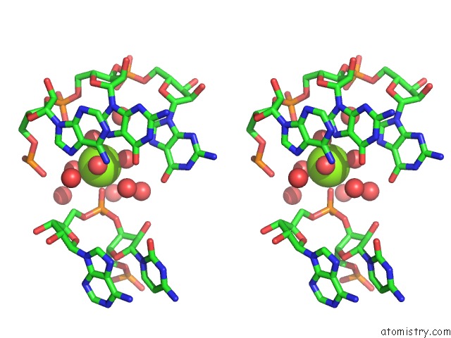
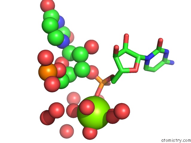
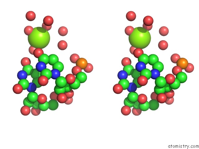
Magnesium binding site 1 out of 6 in 1f27
Go back to
Magnesium binding site 1 out
of 6 in the Crystal Structure of A Biotin-Binding Rna Pseudoknot
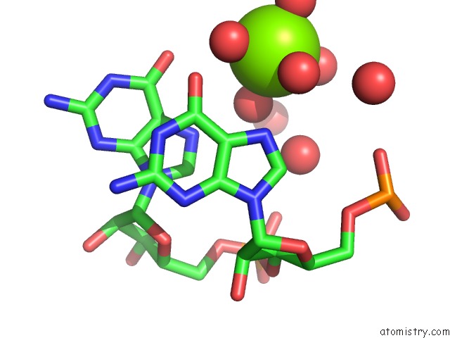
Mono view
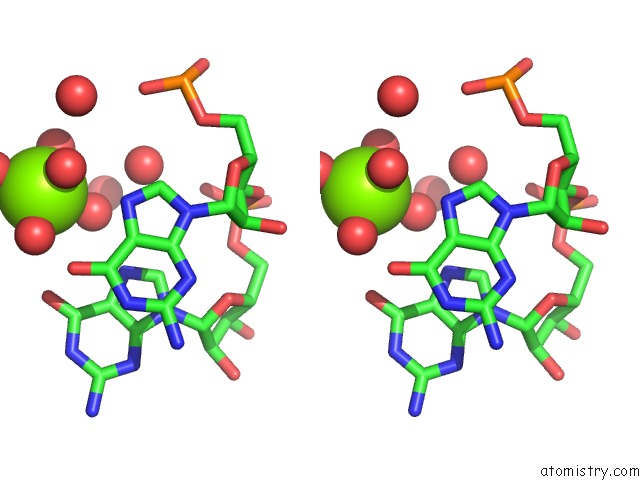
Stereo pair view
Mono view
Stereo pair view
A full contact list of Magnesium with other atoms in the Mg binding
site number 1 of Crystal Structure of A Biotin-Binding Rna Pseudoknot within 5.0Å range:
|
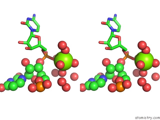
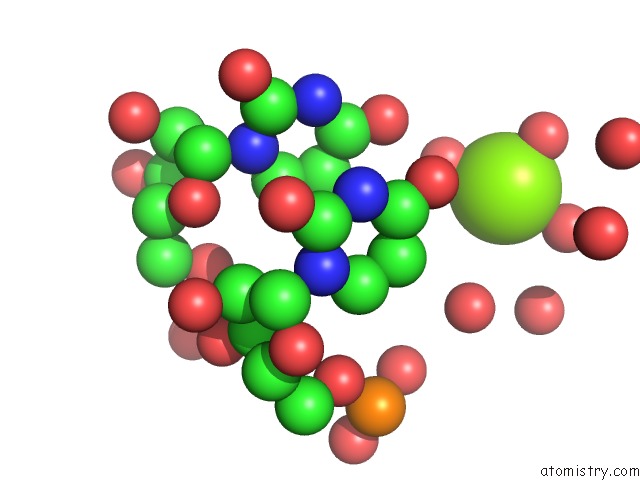
Magnesium binding site 2 out of 6 in 1f27
Go back to
Magnesium binding site 2 out
of 6 in the Crystal Structure of A Biotin-Binding Rna Pseudoknot
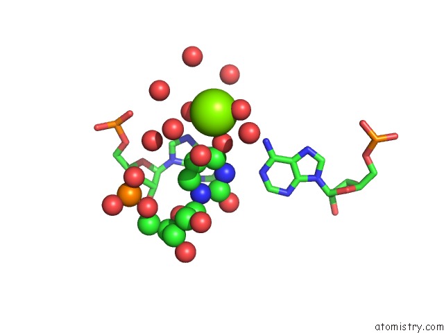
Mono view
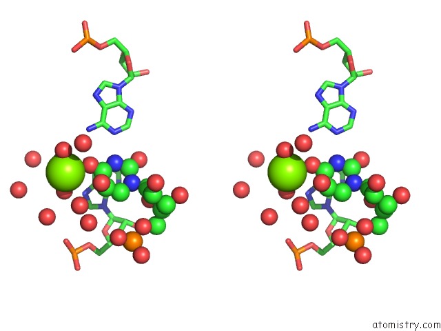
Stereo pair view
Mono view
Stereo pair view
A full contact list of Magnesium with other atoms in the Mg binding
site number 2 of Crystal Structure of A Biotin-Binding Rna Pseudoknot within 5.0Å range:
|
Magnesium binding site 3 out of 6 in 1f27
Go back to
Magnesium binding site 3 out
of 6 in the Crystal Structure of A Biotin-Binding Rna Pseudoknot

Mono view
Stereo pair view
Mono view
Stereo pair view
A full contact list of Magnesium with other atoms in the Mg binding
site number 3 of Crystal Structure of A Biotin-Binding Rna Pseudoknot within 5.0Å range:
|
Magnesium binding site 4 out of 6 in 1f27
Go back to
Magnesium binding site 4 out
of 6 in the Crystal Structure of A Biotin-Binding Rna Pseudoknot
Mono view
Stereo pair view
Mono view
Stereo pair view
A full contact list of Magnesium with other atoms in the Mg binding
site number 4 of Crystal Structure of A Biotin-Binding Rna Pseudoknot within 5.0Å range:
|
Magnesium binding site 5 out of 6 in 1f27
Go back to
Magnesium binding site 5 out
of 6 in the Crystal Structure of A Biotin-Binding Rna Pseudoknot
Mono view
Stereo pair view
Mono view
Stereo pair view
A full contact list of Magnesium with other atoms in the Mg binding
site number 5 of Crystal Structure of A Biotin-Binding Rna Pseudoknot within 5.0Å range:
|
Magnesium binding site 6 out of 6 in 1f27
Go back to
Magnesium binding site 6 out
of 6 in the Crystal Structure of A Biotin-Binding Rna Pseudoknot

Mono view

Stereo pair view
Mono view
Stereo pair view
A full contact list of Magnesium with other atoms in the Mg binding
site number 6 of Crystal Structure of A Biotin-Binding Rna Pseudoknot within 5.0Å range:
|
Reference:
J.Nix,
D.Sussman,
C.Wilson.
The 1.3 A Crystal Structure of A Biotin-Binding Pseudoknot and the Basis For Rna Molecular Recognition. J.Mol.Biol. V. 296 1235 2000.
ISSN: ISSN 0022-2836
PubMed: 10698630
DOI: 10.1006/JMBI.2000.3539
Page generated: Tue Aug 13 03:06:11 2024
ISSN: ISSN 0022-2836
PubMed: 10698630
DOI: 10.1006/JMBI.2000.3539
Last articles
Zn in 9J0NZn in 9J0O
Zn in 9J0P
Zn in 9FJX
Zn in 9EKB
Zn in 9C0F
Zn in 9CAH
Zn in 9CH0
Zn in 9CH3
Zn in 9CH1