Magnesium »
PDB 1eo3-1f6t »
1f3f »
Magnesium in PDB 1f3f: Structure of the H122G Nucleoside Diphosphate Kinase / D4T- Triphosphate.Mg Complex
Enzymatic activity of Structure of the H122G Nucleoside Diphosphate Kinase / D4T- Triphosphate.Mg Complex
All present enzymatic activity of Structure of the H122G Nucleoside Diphosphate Kinase / D4T- Triphosphate.Mg Complex:
2.7.4.6;
2.7.4.6;
Protein crystallography data
The structure of Structure of the H122G Nucleoside Diphosphate Kinase / D4T- Triphosphate.Mg Complex, PDB code: 1f3f
was solved by
P.Meyer,
B.Schneider,
S.Sarfati,
D.Deville-Bonne,
C.Guerreiro,
J.Boretto,
J.Janin,
M.Veron,
B.Canard,
with X-Ray Crystallography technique. A brief refinement statistics is given in the table below:
| Resolution Low / High (Å) | 29.73 / 1.85 |
| Space group | P 31 2 1 |
| Cell size a, b, c (Å), α, β, γ (°) | 70.018, 70.018, 152.098, 90.00, 90.00, 120.00 |
| R / Rfree (%) | 20.3 / 23.4 |
Magnesium Binding Sites:
The binding sites of Magnesium atom in the Structure of the H122G Nucleoside Diphosphate Kinase / D4T- Triphosphate.Mg Complex
(pdb code 1f3f). This binding sites where shown within
5.0 Angstroms radius around Magnesium atom.
In total 3 binding sites of Magnesium where determined in the Structure of the H122G Nucleoside Diphosphate Kinase / D4T- Triphosphate.Mg Complex, PDB code: 1f3f:
Jump to Magnesium binding site number: 1; 2; 3;
In total 3 binding sites of Magnesium where determined in the Structure of the H122G Nucleoside Diphosphate Kinase / D4T- Triphosphate.Mg Complex, PDB code: 1f3f:
Jump to Magnesium binding site number: 1; 2; 3;
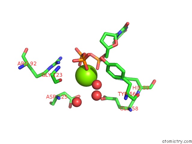
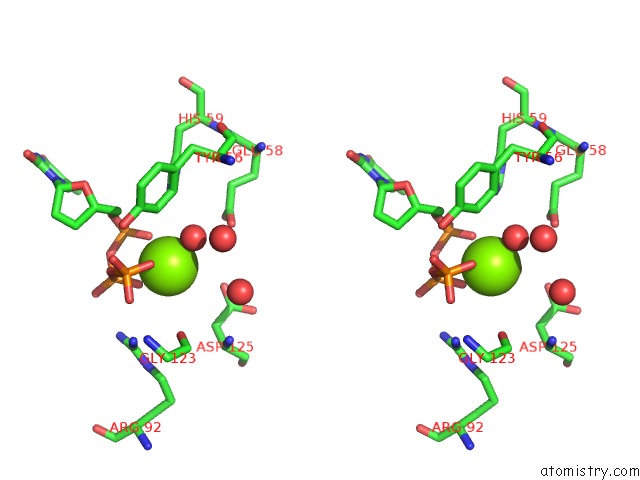
Magnesium binding site 1 out of 3 in 1f3f
Go back to
Magnesium binding site 1 out
of 3 in the Structure of the H122G Nucleoside Diphosphate Kinase / D4T- Triphosphate.Mg Complex
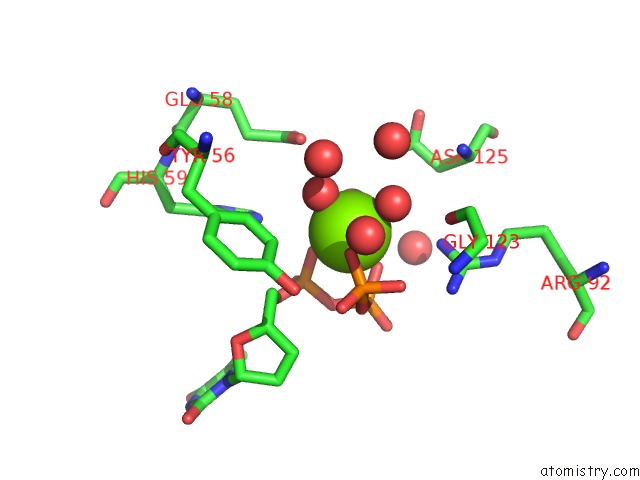
Mono view
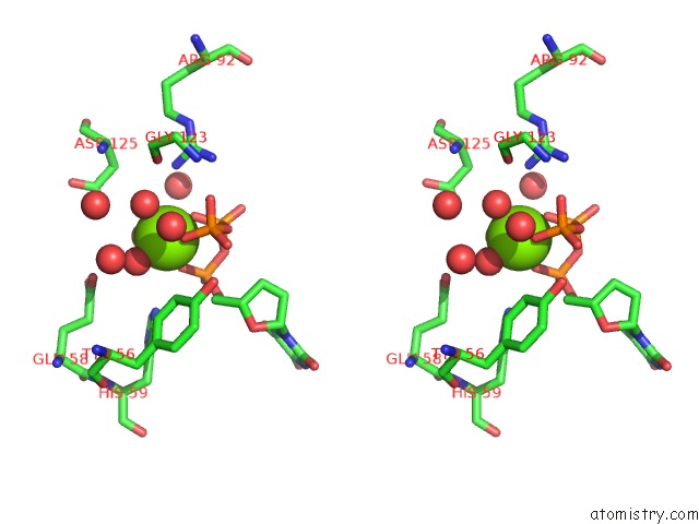
Stereo pair view
Mono view
Stereo pair view
A full contact list of Magnesium with other atoms in the Mg binding
site number 1 of Structure of the H122G Nucleoside Diphosphate Kinase / D4T- Triphosphate.Mg Complex within 5.0Å range:
|
Magnesium binding site 2 out of 3 in 1f3f
Go back to
Magnesium binding site 2 out
of 3 in the Structure of the H122G Nucleoside Diphosphate Kinase / D4T- Triphosphate.Mg Complex
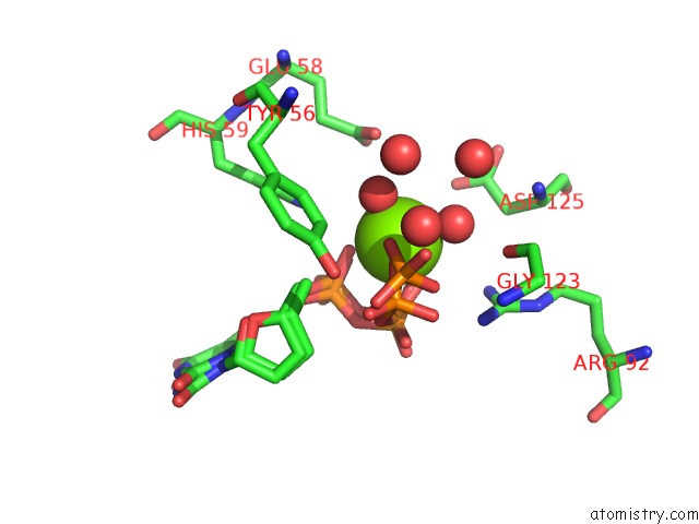
Mono view
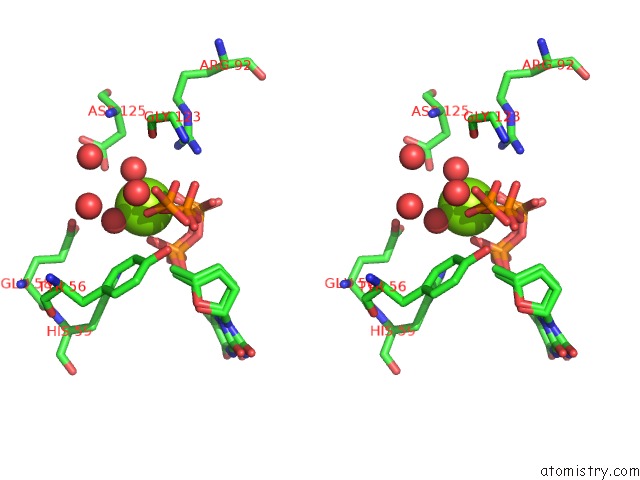
Stereo pair view
Mono view
Stereo pair view
A full contact list of Magnesium with other atoms in the Mg binding
site number 2 of Structure of the H122G Nucleoside Diphosphate Kinase / D4T- Triphosphate.Mg Complex within 5.0Å range:
|
Magnesium binding site 3 out of 3 in 1f3f
Go back to
Magnesium binding site 3 out
of 3 in the Structure of the H122G Nucleoside Diphosphate Kinase / D4T- Triphosphate.Mg Complex
Mono view
Stereo pair view
Mono view
Stereo pair view
A full contact list of Magnesium with other atoms in the Mg binding
site number 3 of Structure of the H122G Nucleoside Diphosphate Kinase / D4T- Triphosphate.Mg Complex within 5.0Å range:
|
Reference:
P.Meyer,
B.Schneider,
S.Sarfati,
D.Deville-Bonne,
C.Guerreiro,
J.Boretto,
J.Janin,
M.Veron,
B.Canard.
Structural Basis For Activation of Alpha-Boranophosphate Nucleotide Analogues Targeting Drug-Resistant Reverse Transcriptase. Embo J. V. 19 3520 2000.
ISSN: ISSN 0261-4189
PubMed: 10899107
DOI: 10.1093/EMBOJ/19.14.3520
Page generated: Tue Aug 13 03:06:20 2024
ISSN: ISSN 0261-4189
PubMed: 10899107
DOI: 10.1093/EMBOJ/19.14.3520
Last articles
Zn in 9MJ5Zn in 9HNW
Zn in 9G0L
Zn in 9FNE
Zn in 9DZN
Zn in 9E0I
Zn in 9D32
Zn in 9DAK
Zn in 8ZXC
Zn in 8ZUF