Magnesium in PDB 1f6n: Crystal Structure Analysis of the Mutant Reaction Center Pro L209-> Tyr From the Photosynthetic Purple Bacterium Rhodobacter Sphaeroides
Protein crystallography data
The structure of Crystal Structure Analysis of the Mutant Reaction Center Pro L209-> Tyr From the Photosynthetic Purple Bacterium Rhodobacter Sphaeroides, PDB code: 1f6n
was solved by
A.Kuglstatter,
U.Ermler,
H.Michel,
L.Baciou,
G.Fritzsch,
with X-Ray Crystallography technique. A brief refinement statistics is given in the table below:
Resolution Low / High (Å)
|
50.00 /
2.80
|
Space group
|
P 31 2 1
|
Cell size a, b, c (Å), α, β, γ (°)
|
141.535,
141.535,
187.913,
90.00,
90.00,
120.00
|
R / Rfree (%)
|
22.1 /
25
|
Other elements in 1f6n:
The structure of Crystal Structure Analysis of the Mutant Reaction Center Pro L209-> Tyr From the Photosynthetic Purple Bacterium Rhodobacter Sphaeroides also contains other interesting chemical elements:
Magnesium Binding Sites:
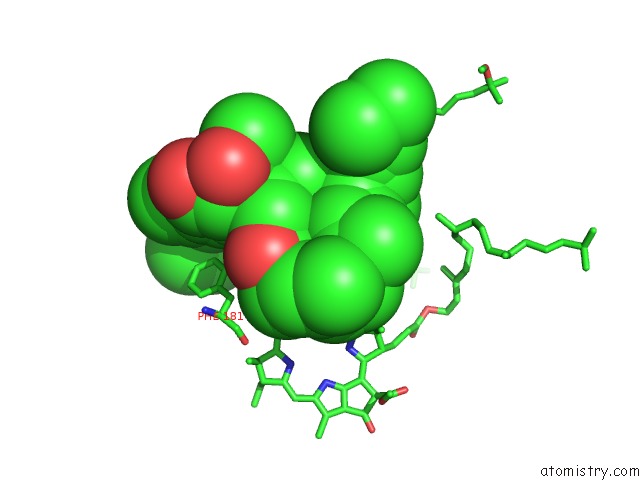
The binding sites of Magnesium atom in the Crystal Structure Analysis of the Mutant Reaction Center Pro L209-> Tyr From the Photosynthetic Purple Bacterium Rhodobacter Sphaeroides
(pdb code 1f6n). This binding sites where shown within
5.0 Angstroms radius around Magnesium atom.
In total 4 binding sites of Magnesium where determined in the
Crystal Structure Analysis of the Mutant Reaction Center Pro L209-> Tyr From the Photosynthetic Purple Bacterium Rhodobacter Sphaeroides, PDB code: 1f6n:
Jump to Magnesium binding site number:
1;
2;
3;
4;
Magnesium binding site 1 out
of 4 in 1f6n
Go back to
Magnesium Binding Sites List in 1f6n
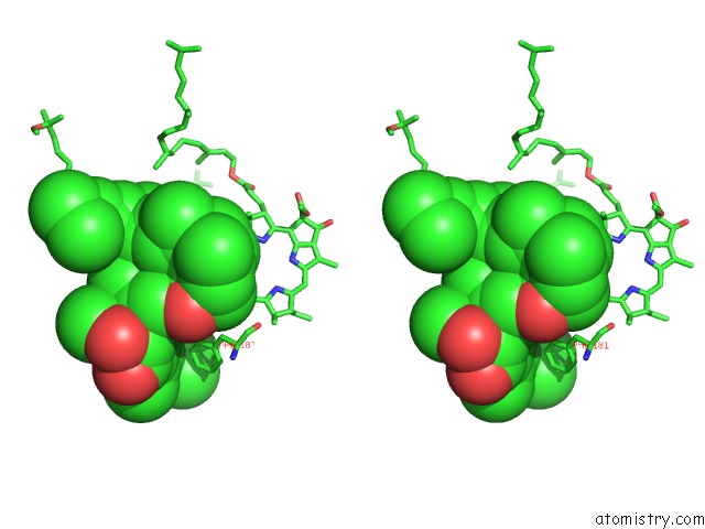
Magnesium binding site 1 out
of 4 in the Crystal Structure Analysis of the Mutant Reaction Center Pro L209-> Tyr From the Photosynthetic Purple Bacterium Rhodobacter Sphaeroides
Mono view
Stereo pair view
|
A full contact list of Magnesium with other atoms in the Mg binding
site number 1 of Crystal Structure Analysis of the Mutant Reaction Center Pro L209-> Tyr From the Photosynthetic Purple Bacterium Rhodobacter Sphaeroides within 5.0Å range:
probe
|
atom
|
residue
|
distance (Å)
|
B
|
Occ
|
L:Mg301
b:60.8
occ:1.00
|
MG
|
L:BCL301
|
0.0
|
60.8
|
1.0
|
ND
|
L:BCL301
|
2.0
|
60.7
|
1.0
|
NB
|
L:BCL301
|
2.1
|
61.2
|
1.0
|
NC
|
L:BCL301
|
2.1
|
60.6
|
1.0
|
NA
|
L:BCL301
|
2.2
|
61.2
|
1.0
|
NE2
|
M:HIS182
|
2.2
|
61.0
|
1.0
|
C4D
|
L:BCL301
|
3.0
|
60.7
|
1.0
|
C4B
|
L:BCL301
|
3.1
|
61.1
|
1.0
|
C1D
|
L:BCL301
|
3.1
|
60.5
|
1.0
|
C1B
|
L:BCL301
|
3.1
|
61.1
|
1.0
|
C1C
|
L:BCL301
|
3.1
|
60.6
|
1.0
|
C4C
|
L:BCL301
|
3.1
|
60.5
|
1.0
|
CE1
|
M:HIS182
|
3.1
|
61.2
|
1.0
|
C4A
|
L:BCL301
|
3.2
|
61.3
|
1.0
|
C1A
|
L:BCL301
|
3.2
|
61.5
|
1.0
|
CD2
|
M:HIS182
|
3.2
|
61.4
|
1.0
|
CHC
|
L:BCL301
|
3.4
|
60.7
|
1.0
|
CHD
|
L:BCL301
|
3.5
|
60.5
|
1.0
|
CHA
|
L:BCL301
|
3.5
|
61.0
|
1.0
|
CHB
|
L:BCL301
|
3.5
|
60.9
|
1.0
|
C3D
|
L:BCL301
|
4.2
|
60.5
|
1.0
|
C2D
|
L:BCL301
|
4.3
|
60.6
|
1.0
|
C2B
|
L:BCL301
|
4.3
|
61.3
|
1.0
|
C3B
|
L:BCL301
|
4.3
|
61.3
|
1.0
|
ND1
|
M:HIS182
|
4.3
|
61.6
|
1.0
|
C2C
|
L:BCL301
|
4.4
|
60.2
|
1.0
|
CG
|
M:HIS182
|
4.4
|
61.7
|
1.0
|
C3C
|
L:BCL301
|
4.4
|
60.2
|
1.0
|
C3A
|
L:BCL301
|
4.5
|
61.7
|
1.0
|
C2A
|
L:BCL301
|
4.5
|
62.3
|
1.0
|
CE2
|
L:PHE181
|
4.8
|
56.7
|
1.0
|
C18
|
M:SPO600
|
4.9
|
74.1
|
1.0
|
C19
|
M:SPO600
|
5.0
|
73.5
|
1.0
|
CBD
|
L:BCL301
|
5.0
|
60.7
|
1.0
|
OBB
|
M:BPH401
|
5.0
|
77.3
|
1.0
|
CAC
|
L:BCL301
|
5.0
|
60.2
|
1.0
|
|
Magnesium binding site 2 out
of 4 in 1f6n
Go back to
Magnesium Binding Sites List in 1f6n
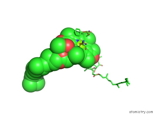
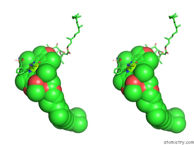
Magnesium binding site 2 out
of 4 in the Crystal Structure Analysis of the Mutant Reaction Center Pro L209-> Tyr From the Photosynthetic Purple Bacterium Rhodobacter Sphaeroides
Mono view
Stereo pair view
|
A full contact list of Magnesium with other atoms in the Mg binding
site number 2 of Crystal Structure Analysis of the Mutant Reaction Center Pro L209-> Tyr From the Photosynthetic Purple Bacterium Rhodobacter Sphaeroides within 5.0Å range:
probe
|
atom
|
residue
|
distance (Å)
|
B
|
Occ
|
L:Mg302
b:52.6
occ:1.00
|
MG
|
L:BCL302
|
0.0
|
52.6
|
1.0
|
ND
|
L:BCL302
|
2.0
|
53.1
|
1.0
|
NB
|
L:BCL302
|
2.1
|
51.4
|
1.0
|
NC
|
L:BCL302
|
2.1
|
52.2
|
1.0
|
NA
|
L:BCL302
|
2.2
|
52.6
|
1.0
|
NE2
|
L:HIS173
|
2.5
|
54.2
|
1.0
|
C4D
|
L:BCL302
|
3.0
|
53.2
|
1.0
|
C1B
|
L:BCL302
|
3.1
|
51.1
|
1.0
|
C4B
|
L:BCL302
|
3.1
|
51.2
|
1.0
|
C1D
|
L:BCL302
|
3.1
|
53.1
|
1.0
|
C1C
|
L:BCL302
|
3.1
|
52.2
|
1.0
|
C4C
|
L:BCL302
|
3.1
|
52.3
|
1.0
|
C4A
|
L:BCL302
|
3.2
|
52.2
|
1.0
|
C1A
|
L:BCL302
|
3.2
|
52.6
|
1.0
|
CE1
|
L:HIS173
|
3.3
|
53.9
|
1.0
|
CHC
|
L:BCL302
|
3.4
|
52.0
|
1.0
|
CHB
|
L:BCL302
|
3.5
|
51.4
|
1.0
|
CD2
|
L:HIS173
|
3.5
|
54.3
|
1.0
|
CHD
|
L:BCL302
|
3.5
|
52.6
|
1.0
|
CHA
|
L:BCL302
|
3.5
|
53.0
|
1.0
|
CBB
|
M:BCL801
|
3.6
|
55.5
|
1.0
|
OBB
|
M:BCL801
|
3.7
|
55.8
|
1.0
|
CAB
|
M:BCL801
|
3.7
|
55.1
|
1.0
|
C3D
|
L:BCL302
|
4.2
|
53.1
|
1.0
|
C2D
|
L:BCL302
|
4.3
|
53.4
|
1.0
|
C2B
|
L:BCL302
|
4.3
|
50.8
|
1.0
|
C3B
|
L:BCL302
|
4.3
|
50.6
|
1.0
|
C2C
|
L:BCL302
|
4.4
|
52.0
|
1.0
|
C3C
|
L:BCL302
|
4.4
|
52.3
|
1.0
|
C3A
|
L:BCL302
|
4.5
|
52.5
|
1.0
|
C3B
|
M:BCL801
|
4.5
|
54.3
|
1.0
|
C2A
|
L:BCL302
|
4.5
|
52.3
|
1.0
|
ND1
|
L:HIS173
|
4.6
|
54.4
|
1.0
|
CG
|
L:HIS173
|
4.6
|
54.7
|
1.0
|
CD1
|
L:PHE167
|
4.8
|
53.9
|
1.0
|
CBD
|
L:BCL302
|
5.0
|
53.4
|
1.0
|
|
Magnesium binding site 3 out
of 4 in 1f6n
Go back to
Magnesium Binding Sites List in 1f6n
Magnesium binding site 3 out
of 4 in the Crystal Structure Analysis of the Mutant Reaction Center Pro L209-> Tyr From the Photosynthetic Purple Bacterium Rhodobacter Sphaeroides
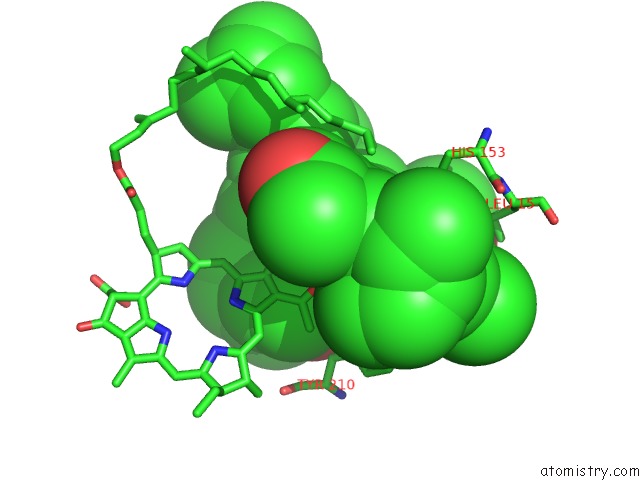
Mono view
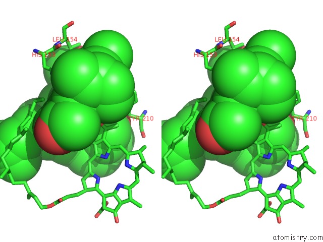
Stereo pair view
|
A full contact list of Magnesium with other atoms in the Mg binding
site number 3 of Crystal Structure Analysis of the Mutant Reaction Center Pro L209-> Tyr From the Photosynthetic Purple Bacterium Rhodobacter Sphaeroides within 5.0Å range:
probe
|
atom
|
residue
|
distance (Å)
|
B
|
Occ
|
L:Mg304
b:40.5
occ:1.00
|
MG
|
L:BCL304
|
0.0
|
40.5
|
1.0
|
ND
|
L:BCL304
|
2.0
|
39.5
|
1.0
|
NC
|
L:BCL304
|
2.0
|
39.1
|
1.0
|
NB
|
L:BCL304
|
2.1
|
40.0
|
1.0
|
NA
|
L:BCL304
|
2.3
|
40.8
|
1.0
|
NE2
|
L:HIS153
|
2.3
|
45.0
|
1.0
|
CHB
|
L:BCL304
|
2.7
|
40.1
|
1.0
|
C4D
|
L:BCL304
|
3.0
|
39.5
|
1.0
|
C1B
|
L:BCL304
|
3.0
|
39.8
|
1.0
|
C1D
|
L:BCL304
|
3.0
|
39.0
|
1.0
|
C4C
|
L:BCL304
|
3.0
|
38.9
|
1.0
|
C1C
|
L:BCL304
|
3.1
|
39.6
|
1.0
|
C4A
|
L:BCL304
|
3.1
|
40.7
|
1.0
|
C4B
|
L:BCL304
|
3.2
|
39.7
|
1.0
|
CE1
|
L:HIS153
|
3.2
|
47.1
|
1.0
|
C1A
|
L:BCL304
|
3.3
|
41.2
|
1.0
|
CD2
|
L:HIS153
|
3.3
|
48.1
|
1.0
|
CHD
|
L:BCL304
|
3.4
|
38.7
|
1.0
|
CHC
|
L:BCL304
|
3.6
|
39.6
|
1.0
|
CHA
|
L:BCL304
|
3.6
|
40.7
|
1.0
|
C3D
|
L:BCL304
|
4.2
|
39.2
|
1.0
|
C2D
|
L:BCL304
|
4.2
|
38.9
|
1.0
|
C2B
|
L:BCL304
|
4.3
|
39.4
|
1.0
|
C3C
|
L:BCL304
|
4.3
|
39.7
|
1.0
|
C2C
|
L:BCL304
|
4.3
|
39.8
|
1.0
|
C3B
|
L:BCL304
|
4.4
|
40.0
|
1.0
|
ND1
|
L:HIS153
|
4.4
|
50.0
|
1.0
|
CBC
|
L:BCL304
|
4.5
|
38.9
|
1.0
|
C3A
|
L:BCL304
|
4.5
|
41.4
|
1.0
|
CG
|
L:HIS153
|
4.5
|
51.0
|
1.0
|
C2A
|
L:BCL304
|
4.6
|
42.1
|
1.0
|
CE2
|
M:TYR210
|
4.6
|
51.8
|
1.0
|
OBB
|
L:BPH402
|
4.7
|
52.3
|
1.0
|
CD2
|
L:LEU154
|
4.7
|
60.0
|
1.0
|
CMC
|
L:BCL304
|
4.9
|
40.5
|
1.0
|
CAC
|
L:BCL304
|
4.9
|
39.6
|
1.0
|
|
Magnesium binding site 4 out
of 4 in 1f6n
Go back to
Magnesium Binding Sites List in 1f6n
Magnesium binding site 4 out
of 4 in the Crystal Structure Analysis of the Mutant Reaction Center Pro L209-> Tyr From the Photosynthetic Purple Bacterium Rhodobacter Sphaeroides
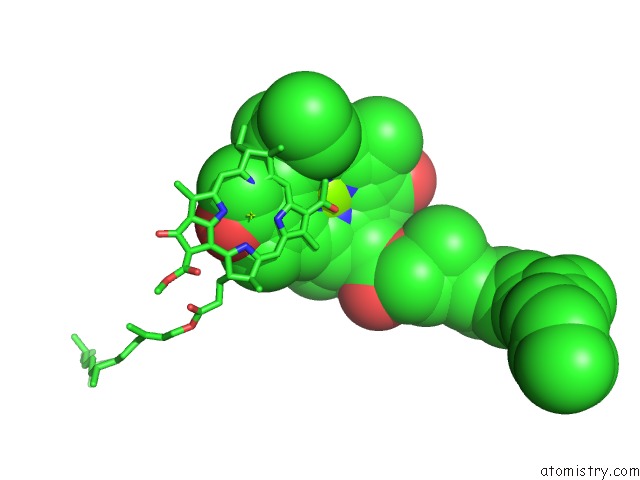
Mono view
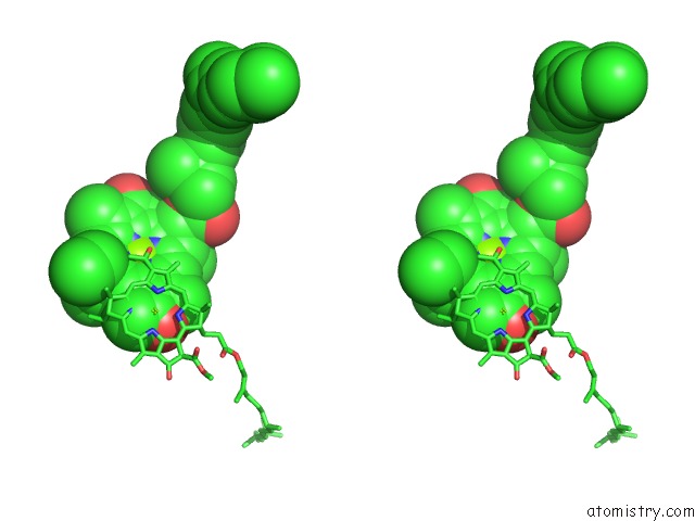
Stereo pair view
|
A full contact list of Magnesium with other atoms in the Mg binding
site number 4 of Crystal Structure Analysis of the Mutant Reaction Center Pro L209-> Tyr From the Photosynthetic Purple Bacterium Rhodobacter Sphaeroides within 5.0Å range:
probe
|
atom
|
residue
|
distance (Å)
|
B
|
Occ
|
M:Mg801
b:53.0
occ:1.00
|
MG
|
M:BCL801
|
0.0
|
53.0
|
1.0
|
ND
|
M:BCL801
|
2.0
|
53.3
|
1.0
|
NB
|
M:BCL801
|
2.1
|
53.3
|
1.0
|
NC
|
M:BCL801
|
2.1
|
53.2
|
1.0
|
NA
|
M:BCL801
|
2.2
|
53.5
|
1.0
|
NE2
|
M:HIS202
|
2.3
|
53.3
|
1.0
|
C4D
|
M:BCL801
|
2.9
|
53.5
|
1.0
|
C1B
|
M:BCL801
|
3.1
|
53.3
|
1.0
|
C4B
|
M:BCL801
|
3.1
|
53.5
|
1.0
|
C1C
|
M:BCL801
|
3.1
|
53.4
|
1.0
|
C1D
|
M:BCL801
|
3.1
|
53.2
|
1.0
|
C4C
|
M:BCL801
|
3.1
|
53.1
|
1.0
|
C4A
|
M:BCL801
|
3.2
|
53.6
|
1.0
|
C1A
|
M:BCL801
|
3.2
|
53.6
|
1.0
|
CE1
|
M:HIS202
|
3.2
|
53.0
|
1.0
|
CD2
|
M:HIS202
|
3.3
|
53.1
|
1.0
|
CHC
|
M:BCL801
|
3.4
|
53.3
|
1.0
|
CHA
|
M:BCL801
|
3.4
|
53.6
|
1.0
|
CHB
|
M:BCL801
|
3.5
|
53.3
|
1.0
|
CHD
|
M:BCL801
|
3.5
|
53.2
|
1.0
|
OBB
|
L:BCL302
|
3.6
|
50.6
|
1.0
|
CAB
|
L:BCL302
|
3.7
|
49.8
|
1.0
|
CBB
|
L:BCL302
|
4.1
|
49.4
|
1.0
|
C3D
|
M:BCL801
|
4.2
|
53.5
|
1.0
|
C2B
|
M:BCL801
|
4.3
|
53.8
|
1.0
|
C3B
|
M:BCL801
|
4.3
|
54.3
|
1.0
|
C2D
|
M:BCL801
|
4.3
|
53.2
|
1.0
|
C2C
|
M:BCL801
|
4.3
|
53.4
|
1.0
|
C3C
|
M:BCL801
|
4.4
|
53.2
|
1.0
|
ND1
|
M:HIS202
|
4.4
|
53.1
|
1.0
|
C3A
|
M:BCL801
|
4.5
|
54.0
|
1.0
|
C2A
|
M:BCL801
|
4.5
|
54.0
|
1.0
|
CG
|
M:HIS202
|
4.5
|
53.3
|
1.0
|
C3B
|
L:BCL302
|
4.5
|
50.6
|
1.0
|
CBD
|
M:BCL801
|
4.9
|
54.0
|
1.0
|
CMC
|
M:BCL801
|
4.9
|
53.3
|
1.0
|
|
Reference:
A.Kuglstatter,
U.Ermler,
H.Michel,
L.Baciou,
G.Fritzsch.
X-Ray Structure Analyses of Photosynthetic Reaction Center Variants From Rhodobacter Sphaeroides: Structural Changes Induced By Point Mutations at Position L209 Modulate Electron and Proton Transfer. Biochemistry V. 40 4253 2001.
ISSN: ISSN 0006-2960
PubMed: 11284681
DOI: 10.1021/BI001589H
Page generated: Tue Aug 13 03:09:12 2024
|