Magnesium »
PDB 1f7d-1fnt »
1f9a »
Magnesium in PDB 1f9a: Crystal Structure Analysis of Nmn Adenylyltransferase From Methanococcus Jannaschii
Protein crystallography data
The structure of Crystal Structure Analysis of Nmn Adenylyltransferase From Methanococcus Jannaschii, PDB code: 1f9a
was solved by
I.D'angelo,
N.Raffaelli,
V.Dabusti,
T.Lorenzi,
G.Magni,
M.Rizzi,
with X-Ray Crystallography technique. A brief refinement statistics is given in the table below:
| Resolution Low / High (Å) | 20.00 / 2.00 |
| Space group | P 1 21 1 |
| Cell size a, b, c (Å), α, β, γ (°) | 78.768, 112.642, 79.869, 90.00, 116.94, 90.00 |
| R / Rfree (%) | 21.5 / 26.4 |
Magnesium Binding Sites:
The binding sites of Magnesium atom in the Crystal Structure Analysis of Nmn Adenylyltransferase From Methanococcus Jannaschii
(pdb code 1f9a). This binding sites where shown within
5.0 Angstroms radius around Magnesium atom.
In total 6 binding sites of Magnesium where determined in the Crystal Structure Analysis of Nmn Adenylyltransferase From Methanococcus Jannaschii, PDB code: 1f9a:
Jump to Magnesium binding site number: 1; 2; 3; 4; 5; 6;
In total 6 binding sites of Magnesium where determined in the Crystal Structure Analysis of Nmn Adenylyltransferase From Methanococcus Jannaschii, PDB code: 1f9a:
Jump to Magnesium binding site number: 1; 2; 3; 4; 5; 6;
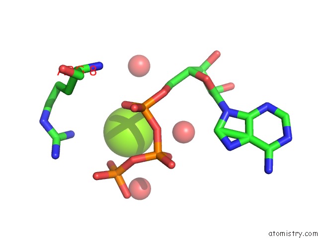
Magnesium binding site 1 out of 6 in 1f9a
Go back to
Magnesium binding site 1 out
of 6 in the Crystal Structure Analysis of Nmn Adenylyltransferase From Methanococcus Jannaschii
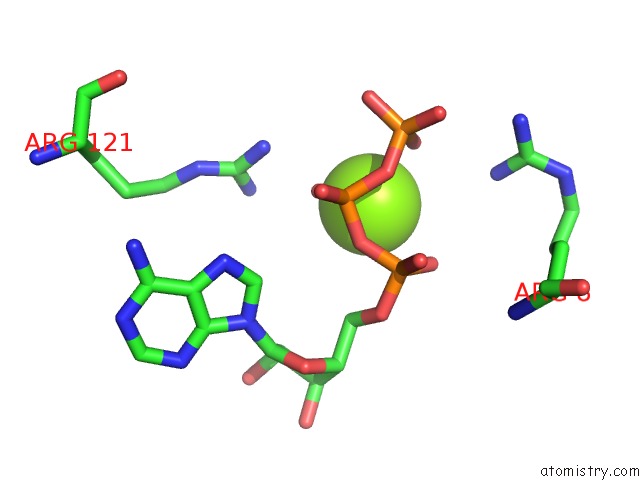
Mono view
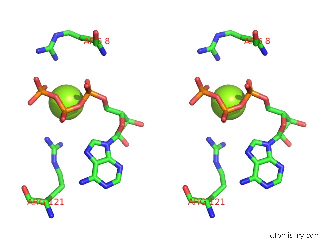
Stereo pair view
Mono view
Stereo pair view
A full contact list of Magnesium with other atoms in the Mg binding
site number 1 of Crystal Structure Analysis of Nmn Adenylyltransferase From Methanococcus Jannaschii within 5.0Å range:
|
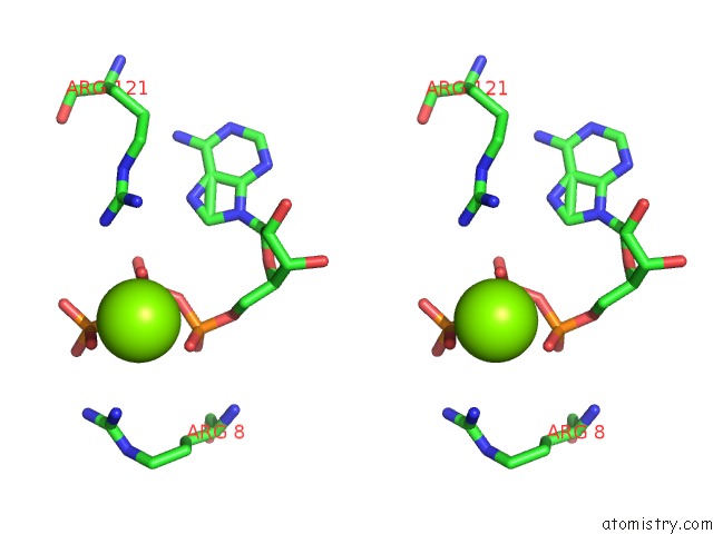
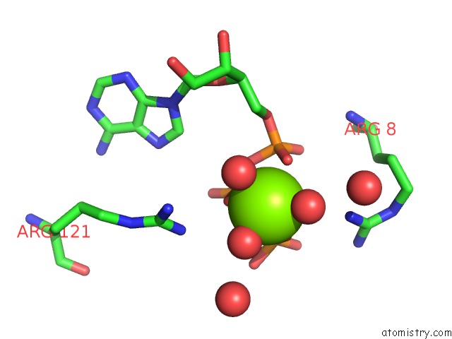
Magnesium binding site 2 out of 6 in 1f9a
Go back to
Magnesium binding site 2 out
of 6 in the Crystal Structure Analysis of Nmn Adenylyltransferase From Methanococcus Jannaschii

Mono view

Stereo pair view
Mono view
Stereo pair view
A full contact list of Magnesium with other atoms in the Mg binding
site number 2 of Crystal Structure Analysis of Nmn Adenylyltransferase From Methanococcus Jannaschii within 5.0Å range:
|
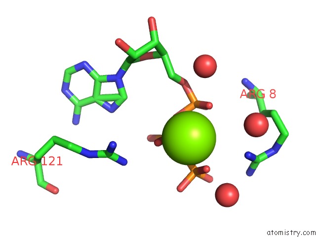
Magnesium binding site 3 out of 6 in 1f9a
Go back to
Magnesium binding site 3 out
of 6 in the Crystal Structure Analysis of Nmn Adenylyltransferase From Methanococcus Jannaschii

Mono view
Stereo pair view
Mono view
Stereo pair view
A full contact list of Magnesium with other atoms in the Mg binding
site number 3 of Crystal Structure Analysis of Nmn Adenylyltransferase From Methanococcus Jannaschii within 5.0Å range:
|
Magnesium binding site 4 out of 6 in 1f9a
Go back to
Magnesium binding site 4 out
of 6 in the Crystal Structure Analysis of Nmn Adenylyltransferase From Methanococcus Jannaschii
Mono view

Stereo pair view
Mono view
Stereo pair view
A full contact list of Magnesium with other atoms in the Mg binding
site number 4 of Crystal Structure Analysis of Nmn Adenylyltransferase From Methanococcus Jannaschii within 5.0Å range:
|
Magnesium binding site 5 out of 6 in 1f9a
Go back to
Magnesium binding site 5 out
of 6 in the Crystal Structure Analysis of Nmn Adenylyltransferase From Methanococcus Jannaschii
Mono view

Stereo pair view
Mono view
Stereo pair view
A full contact list of Magnesium with other atoms in the Mg binding
site number 5 of Crystal Structure Analysis of Nmn Adenylyltransferase From Methanococcus Jannaschii within 5.0Å range:
|
Magnesium binding site 6 out of 6 in 1f9a
Go back to
Magnesium binding site 6 out
of 6 in the Crystal Structure Analysis of Nmn Adenylyltransferase From Methanococcus Jannaschii
Mono view

Stereo pair view
Mono view
Stereo pair view
A full contact list of Magnesium with other atoms in the Mg binding
site number 6 of Crystal Structure Analysis of Nmn Adenylyltransferase From Methanococcus Jannaschii within 5.0Å range:
|
Reference:
I.D'angelo,
N.Raffaelli,
V.Dabusti,
T.Lorenzi,
G.Magni,
M.Rizzi.
Structure of Nicotinamide Mononucleotide Adenylyltransferase: A Key Enzyme in Nad(+) Biosynthesis. Structure Fold.Des. V. 8 993 2000.
ISSN: ISSN 0969-2126
PubMed: 10986466
DOI: 10.1016/S0969-2126(00)00190-8
Page generated: Tue Aug 13 03:12:44 2024
ISSN: ISSN 0969-2126
PubMed: 10986466
DOI: 10.1016/S0969-2126(00)00190-8
Last articles
Zn in 9MJ5Zn in 9HNW
Zn in 9G0L
Zn in 9FNE
Zn in 9DZN
Zn in 9E0I
Zn in 9D32
Zn in 9DAK
Zn in 8ZXC
Zn in 8ZUF