Magnesium »
PDB 1fp6-1g7t »
1g3u »
Magnesium in PDB 1g3u: Crystal Structure of Mycobacterium Tuberculosis Thymidylate Kinase Complexed with Thymidine Monophosphate (Tmp)
Enzymatic activity of Crystal Structure of Mycobacterium Tuberculosis Thymidylate Kinase Complexed with Thymidine Monophosphate (Tmp)
All present enzymatic activity of Crystal Structure of Mycobacterium Tuberculosis Thymidylate Kinase Complexed with Thymidine Monophosphate (Tmp):
2.7.4.9;
2.7.4.9;
Protein crystallography data
The structure of Crystal Structure of Mycobacterium Tuberculosis Thymidylate Kinase Complexed with Thymidine Monophosphate (Tmp), PDB code: 1g3u
was solved by
I.Li De La Sierra,
H.Munier-Lehmann,
A.M.Gilles,
O.Barzu,
M.Delarue,
with X-Ray Crystallography technique. A brief refinement statistics is given in the table below:
| Resolution Low / High (Å) | 20.00 / 1.95 |
| Space group | P 65 2 2 |
| Cell size a, b, c (Å), α, β, γ (°) | 76.622, 76.622, 134.378, 90.00, 90.00, 120.00 |
| R / Rfree (%) | 21.6 / 25 |
Magnesium Binding Sites:
The binding sites of Magnesium atom in the Crystal Structure of Mycobacterium Tuberculosis Thymidylate Kinase Complexed with Thymidine Monophosphate (Tmp)
(pdb code 1g3u). This binding sites where shown within
5.0 Angstroms radius around Magnesium atom.
In total only one binding site of Magnesium was determined in the Crystal Structure of Mycobacterium Tuberculosis Thymidylate Kinase Complexed with Thymidine Monophosphate (Tmp), PDB code: 1g3u:
In total only one binding site of Magnesium was determined in the Crystal Structure of Mycobacterium Tuberculosis Thymidylate Kinase Complexed with Thymidine Monophosphate (Tmp), PDB code: 1g3u:
Magnesium binding site 1 out of 1 in 1g3u
Go back to
Magnesium binding site 1 out
of 1 in the Crystal Structure of Mycobacterium Tuberculosis Thymidylate Kinase Complexed with Thymidine Monophosphate (Tmp)
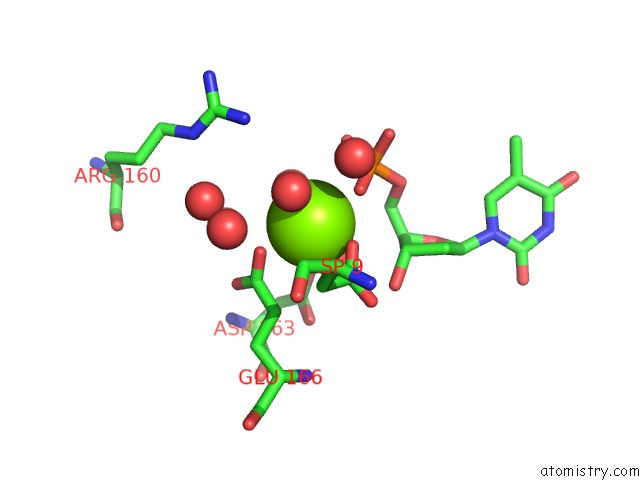
Mono view
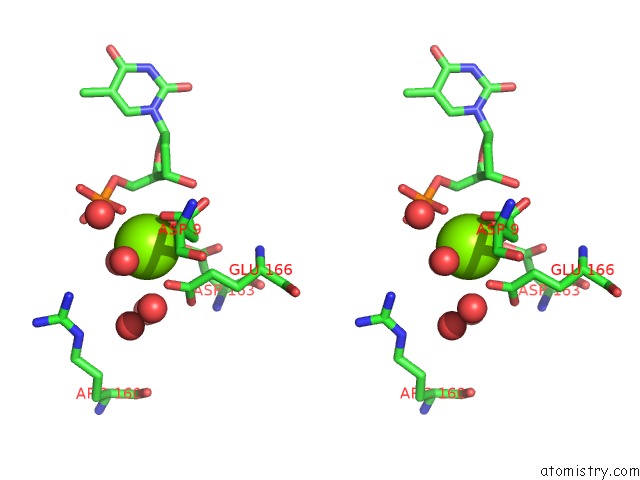
Stereo pair view
Mono view
Stereo pair view
A full contact list of Magnesium with other atoms in the Mg binding
site number 1 of Crystal Structure of Mycobacterium Tuberculosis Thymidylate Kinase Complexed with Thymidine Monophosphate (Tmp) within 5.0Å range:
|
Reference:
I.Li De La Sierra,
H.Munier-Lehmann,
A.M.Gilles,
O.Barzu,
M.Delarue.
X-Ray Structure of Tmp Kinase From Mycobacterium Tuberculosis Complexed with Tmp at 1.95 A Resolution. J.Mol.Biol. V. 311 87 2001.
ISSN: ISSN 0022-2836
PubMed: 11469859
DOI: 10.1006/JMBI.2001.4843
Page generated: Tue Aug 13 03:37:16 2024
ISSN: ISSN 0022-2836
PubMed: 11469859
DOI: 10.1006/JMBI.2001.4843
Last articles
Zn in 9JYWZn in 9IR4
Zn in 9IR3
Zn in 9GMX
Zn in 9GMW
Zn in 9JEJ
Zn in 9ERF
Zn in 9ERE
Zn in 9EGV
Zn in 9EGW